Synthesis of Cu(II), Ni(II), Co(II), and Mn(II) Complexes with Ciprofloxacin and Their Evaluation of Antimicrobial, Antioxidant and Anti-Tubercular Activity ()
1. Introduction
The development of sensitive chemosensors is an active field of research in recent years because of their potential application in clinical biochemistry as well as analytical chemistry and environmental science [1-3]. Coumarins are wide spread in nature, also the biological properties of different coumarins and their derivatives are well known-they include anticoagulant, antiproliferative, antimicrobial, spasmolytic, antitumor, antioxidant, etc. activities [4-6].
The spectrum of activity of these compounds has become increasingly broad, especially since the introduction of a fluorine atom at position 6 (fluoroquinolones) [7]. In 2002, Turel [8] reviewed the synthesis, physico-chemical properties, and structural characteristics of several binary and ternary fluoroquinolone compounds, together with their biological activity. Ciprofloxacin [CF, 1-cyclopro-pyl-6-fluoro-1,4-dihydro-4-oxo-7-(1-piperaz-inil) -3-quinolone car-boxylic acid], is a second generation fluoroquinolone that was synthesized for first time in 1987 [7]. A well known antibacterial drug with a wide spectrum of activity, it is extremely useful for the treatment of a variety of infections [8,9]. Quinolones are a group of synthetic antibacterial agents now in clinical use already for over thirty years and ciprofloxacin is one of the widely used representatives [10,11]. The interactions of quinolones and metal ions have been thoroughly studied especially due to the interesting biological and chemical properties Ciprofloxacin can usually act as a bidentate ligand through the pyridone oxygen and one carboxylate oxygen. In the literature, diverse transition metal complexes of ciprofloxacin have been structurally characterized.
It is well known that metal ions present in complexes accelerate the drug action and the efficacy of the organic therapeutic agents [12]. The pharmacological efficiencies of metal complexes depend on the nature of the metal ions and the ligands [13]. It is declared in the literature that different ligands and different complexes synthesized from same ligands with different metal ions possess different biological properties [12,14,15]. So, there is an increasing requirement for the discovery of new compounds having antimicrobial, antioxidant and anti tubercular activities. The newly prepared compounds may be more effective than known others in terms of their biological activities and possibly display their efficiencies with a distinct mechanism from those of well known, Also we describes the synthesis of Cu(II), Ni(II), Co(II) and Mn(II) complexes from bromocoumarin and ciprofloxacin as ligand. For characterization of the compounds, following spectroscopic and analytical techniques were employed: IR, NMR, TGA and elemental analyses.
2. Experimental
2.1. Materials
All reagents were of analytical reagent (AR) grade purchased commercially from Spectro chem. Ltd., Mumbai-India and used without further purification. Solvents employed were distilled, purified and dried by standard procedures prior to use [15]. Clioquinol was purchased from Agro Chemical Division, Atul Ltd., Valsad-India. The metal nitrates used were in hydrated form.
2.2. Physical Measurements
All reactions were monitored by thin-layer chromatography (TLC on alluminium plates coated with silica gel 60 F254, 0.25 mm thickness, E. Merck, Mumbai-India) and detection of the components were measured under UV light or explore in Iodine chamber. Carbon, hydrogen and nitrogen were estimated by elemental analyzer PerkinElmer, USA 2400-II CHN analyzer. Metal ion analyses was carry out by the dissolution of solid complex in hot concentrated nitric acid, further diluting with distilled water and filtered to remove the precipitated organic ligands. Remaining solution was neutralized with ammonia solution and the metal ions were titrated against EDTA. 1H and 13C NMR measurements were carried out on Advance-II 400 Bruker NMR spectrometer, SAIF, Chandigarh. The chemical shifts were measured with respect to TMS which used as internal standard and DMSO-d6 used as solvent. Infrared spectra of solids were recorded in the region 400 - 4000 cm−1 on a Nicolet Impact 400D Fourier-Transform Infrared Spectrophotometer using KBr pellets. Melting point of the ligands and metal complexes were measured by open capillary tube method. Thermal decomposition (TG) analysis was obtained by a model Diamond TGA, PerkinElmer, USAThe experiments were performed in N2 atmosphere at a heating rate of 20˚C min−1 in the temperature range 30˚C - 800˚C.
2.3. Synthesis of 6-Bromo-3-acetyl Coumarin
6-Bromo-3-acetyl coumarin was prepared according to the reported method [16]. A mixture of 6-bromo salicylaldhyde (12.2 g, 0.1 mol), ethylacetoacetate (13.0 g, 0.1 mol) and 3 to 4 drop piperidine were stirred for 10 mins. at room temperature in a 100 ml round bottom flask. After 10 mins. it was heated for 30 mins in water bath. A yellow solid obtained was taken out and washed with cold ether. It was recrystallized from chloroform-hexane. Yield: 92%; M.p.119.5˚C.
2.4. Synthesis of 2.4.1 Synthesis of 6-Bromo-3-cinnamoyl-2H-chromen-2-one (L)
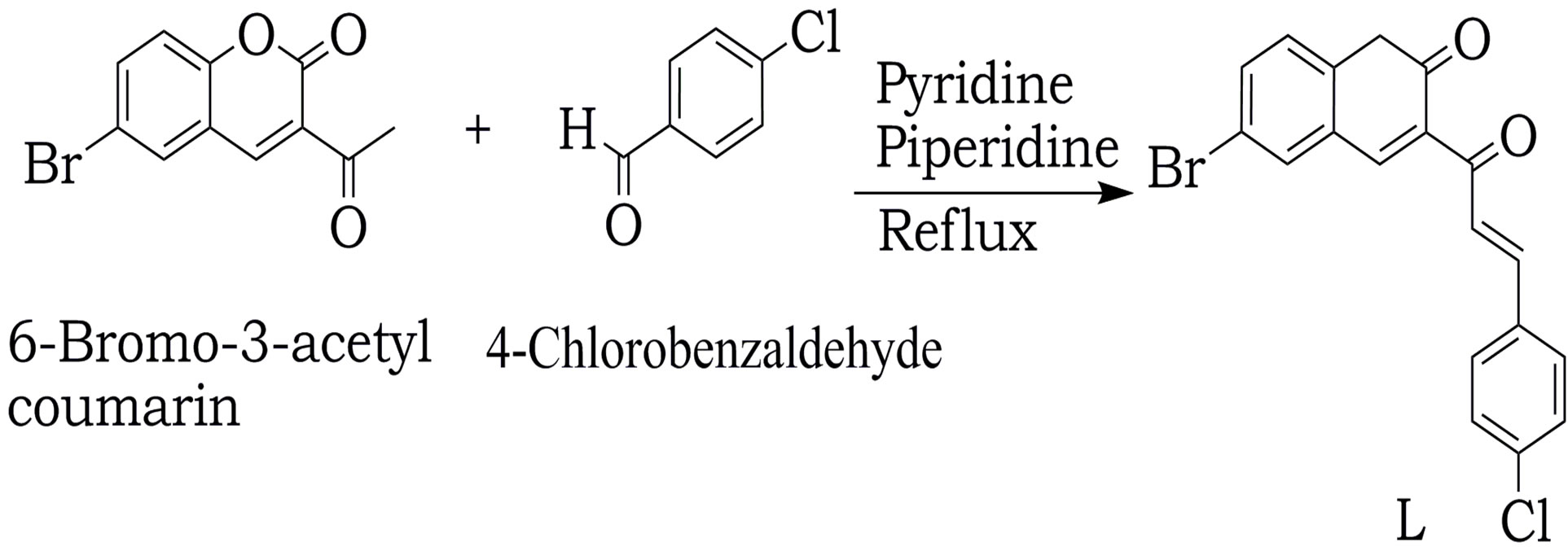
The neutral bidentate ligands were synthesized using Claisen-Schmidt condensation [17]. General procedure for synthesis of the ligands (L) is shown in Scheme 1.
The ligands were characterized using elemental analysis. In a 100 ml round bottom flask 6-bromo 3-acetyl coumarin (0.01 mol, 1.88 g) and 4-Chloro benzaldehyde (0.015 mol) were taken in 15 ml of pyridine. Catalytic amount of piperidine (1.0 ml) was added and the reaction mixture was stirred for 10 min at room temperature. After clear solution obtained, the reaction mixture was refluxed on oil bath. Completion of reaction was checked by TLC using mobile phase Ethyl acetate:Hexane (7:3). After the completion of reaction, subsequently it was allowed to room temperature. Afterwards it was pour into ice-cold water and adjust the pH 4 - 5 using diluted HCl. A solid product separated out was filtered off, later on washed with cold ethanol and dried in air. It was recrystallized from ethanol. Yield: 76%, m.p.: 164˚C - 165˚C. FT-IR (KBr, cm−1): ν (C=O, α, β-unsaturated ketone) 1621, ν (C=O, lactone carbonyl of coumarin) 1738, 1031, (p-substituted C-Cl). 1H NMR (DMSO-d6 400 MHz) δ: 6.81 (1H, d, CH=CH-protons), 7.34 (2H, d, CH=CHprotons), 7.58 (2H, d, CH=CH-protons), 7.78 - 8.08 (3H, m, three aromatic protons), 8.12 (1H, d, CH=CH-protons), 8.59 (1H, s, C4-H). 13C NMR (DMSO-d6 100 MHz) δ: 118.7, 119.4, 124.6, 125.1, 128.2, 129.7, 129.3, 133.1, 133.8, 130.2, 134.9, 142.6 (12 different types of aromatic carbons), 147.6(C-4), 152.3(C-9), 159.7(C=O, lactone carbonyl of coumarin), 183.8(C=O, α, β-unsaturated ketone). MS (ESI) m/z 390.0 [M+H]+, 392.0 [M+H]2+, 394.0 [M+H]4+; Elemental analysis found (%): C, 55.27; H, 2.41; Calculated for C18H10BrClO3 (389.63): C, 55.49; H, 2.59 (Figures 1 and 2).
2.5. Synthesis of Metal Complexes
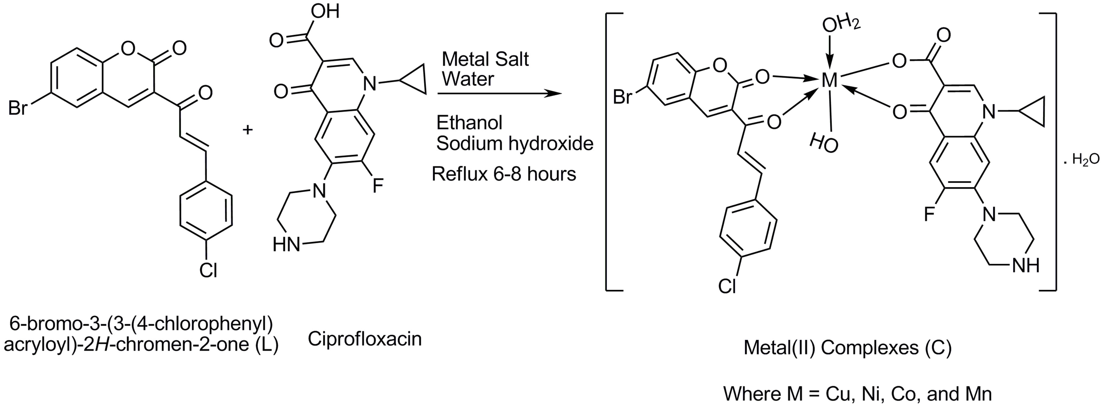
An aqueous solution of Cu(NO3)23H2O salt (10 mmol) was added into ethanolic solution of ligand (L) (10 mmol) and subsequently an ethanolic solution of ciprofloxacin (10 mmol) was added with continuous stirring. Then the pH was adjusted in between 4.5 - 6.0 by addition of diluted NH4OH solution. The resulting solution was refluxed for 5 hrs and then heated over a steam bath to evaporate up to half of the volume. The reaction mixture was kept overnight at room temperature. A fine coloured crystalline product was obtained. The obtained product
Scheme 1. Procedure for synthesis of ligands (L)
was washed with ether and dried over vacuum desiccators. Complexes Ni(II), Co(II) and Mn(II) was prepared according to same method and their physicochemical parameters are summarized in Table 1. The synthetic protocol of complexes is shown in Scheme 2.
2.6. Antimicrobial Activity
The synthesized ligand and corresponding metal(II) complexes were screened in vitro for their antibacterial activity against two Gram(+ve) Streptococcus pyogenes, Bacillus subtilis and two Gram(−ve) Escherichia coli, Pseudomonas aeruginosa, where antifungal against Candida albicans and Aspergillus niger using the brothdilution method [18]. All the ATCC culture was collected from institute of microbial technology, Bangalore. 2% Luria broth solution was prepared in distilled water while, pH of the solution was adjusted to 7.4 ± 0.2 at room temperature and sterilized by autoclaving at 15 lb pressure for 25 mins. The tested bacterial and fungal strains were prepared in the luria broth and incubated at 37˚C and 200 rpm in an orbital incubator for overnight. Sample solutions were prepared in DMSO for concentration 200, 150, 100, 50, 25, 12.5, 6.25 and 3.125, µg/ml. The standard drug solution of Streptomycin (antibacterial drug) and Nystatin (antifungal drug) were prepared in DMSO. Serial broth micro dilution was adopted as a reference method. 10 µl solution of test compound was inoculated in 5 ml luria broth for each concentration respectively and additionally one test tubes was kept as control. Each of the test tubes was inoculated with a suspension of standard microorganism to be tested and incubated at 35˚C for 24 hrs. At the end of the incubation period, the tubes were examined for the turbidity. Turbidity in the test tubes indicated that microorganism growth has not inhibited by the antibiotic contained in the medium at the test concentration. The antimicrobial activity tests were run in triplicate.
2.7. Anti-Tubercular Activity
Test compounds were evaluated for in vitro antimycobacterial activity. The MICs were determined and interpreted for M. tuberculosis H37Rv according to the procedure of the approved microdilution reference method of antimicrobial susceptibility testing [19]. Compounds were taken at concentrations of 100, 50, 25, 12.5 6.25 and 3.125 µg/ml in 2% DMF. M. tuberculosis H37Rv

Table 1. Analytical and physical parameters of complex.
Scheme 2. General procedure for synthesis of complexe (C)
strain was used in Middle brook 7H-9 broth which was inoculated with standard as well as test compounds and incubated at 37˚C for 4 weeks. The bottles were inspected for growth twice a week for a period of 3 weeks. Readings were taken at the end of fourth week. The appearance of turbidity was considered as bacterial growth and indicates resistance to the compound. The growth was confirmed by making a smear from each bottle and performing a Zn stain. Test compounds were compared to reference drugs Ethambutol (MIC = 3.25 µg/ml) and the antimicrobial and anti-tubercular activity tests were run in triplicate.
2.8. Antioxidant Studies
Ferric reducing antioxidant power (FRAP) was measured by a modified method of Benzie and Strain [20]. The antioxidant potentials of the compounds were estimated as their power to reduce the TPTZ-Fe(III) complex to TPTZ-Fe(II) complex (FRAP assay), which is simple, fast, and reproducible. FRAP working solution was prepared by mixing a 25.0 ml, 10 mm TPTZ solution in 40 mm HCl, 20 mm FeCl3.6H2O and 25 ml, 0.3 M acetate buffer at pH 3.6. A mixture of 40.0 ml, 0.5 mm sample solution and 1.2 ml FRAP reagent was incubated at 37˚C for 15 mins. Absorbance of intensive blue colour [Fe(II)-TPTZ] complex was measured at 593 nm. The ascorbic acid was used as a standard antioxidant compound. The results are expressed as ascorbic equivalent (mmol/100 g of dried compound). All the tests were run in triplicate and are expressed as the mean and standard deviation (SD).
3. Result and Discussion
The synthesized complexes were characterized by elemental analysis and FTIR. The metal ion in their complexes was determined after mineralization. The metal content in chemical analysis was estimated by complexometrically [21]. However, ligand and its complexes have been screened for their in vitro antimicrobial antioxidant and anti-tubercular activities, while geometry of the complexes was confirmed from electronic spectra and TGA.
3.1. Elemental Analysis
The analytical and physiochemical data of the complexes are summarized in Table 1. The experimental data were in very good agreement with the calculated ones. The complexes were colored, insoluble in water and commonly organic solvents while soluble in DMSO as well as stable in air.
3.2. FT-IR Spectra
The analysis of the FT-IR spectra of both ligands and complex provided information on the coordination mode between the ligands and the metal ion IR Spectra. The IR Spectra of ligand (L) and its Co(II) complex are given in Figures 3 and 4 respectively, while the IR spectral data of all complexes are summarized in Table 2. The infrared spectra of fluoroquinolones are quite complex due to the presence of the numerous functional groups in the molecules, therefore their interpretation is based on the most typical vibrations being the most important region in the IR spectra of fluoroquinolones between ≈1800 cm−1 and ≈1300 cm−1 [22]. Spectra of the mixed-ligand Cu(II) complexes reveals that a broad band in the region ≈3420 - 3460 cm−1 due to stretching vibration of OH group. The ν (C=O) stretching vibration band appears at ≈1708 cm–1 in the spectra of ciprofloxacin, and the complexes show this band at ≈1628 cm–1; this band shifted towards lower energy, suggesting that coordination occurs through the pyridone oxygen atom [23]. The strong absorption bands obtained at ≈1625 and ≈1380 cm–1 in ciprofloxacin are observed at ≈1570 - 1580 and ≈1345 - 1375 cm–1 for ν(COO)a and ν (COO)s in the complexes, respectively; in the present case the separation frequency Δν > 200 cm–1 (Δν = ν (COO)a − ν (COO)s ), suggesting unidentate binding of
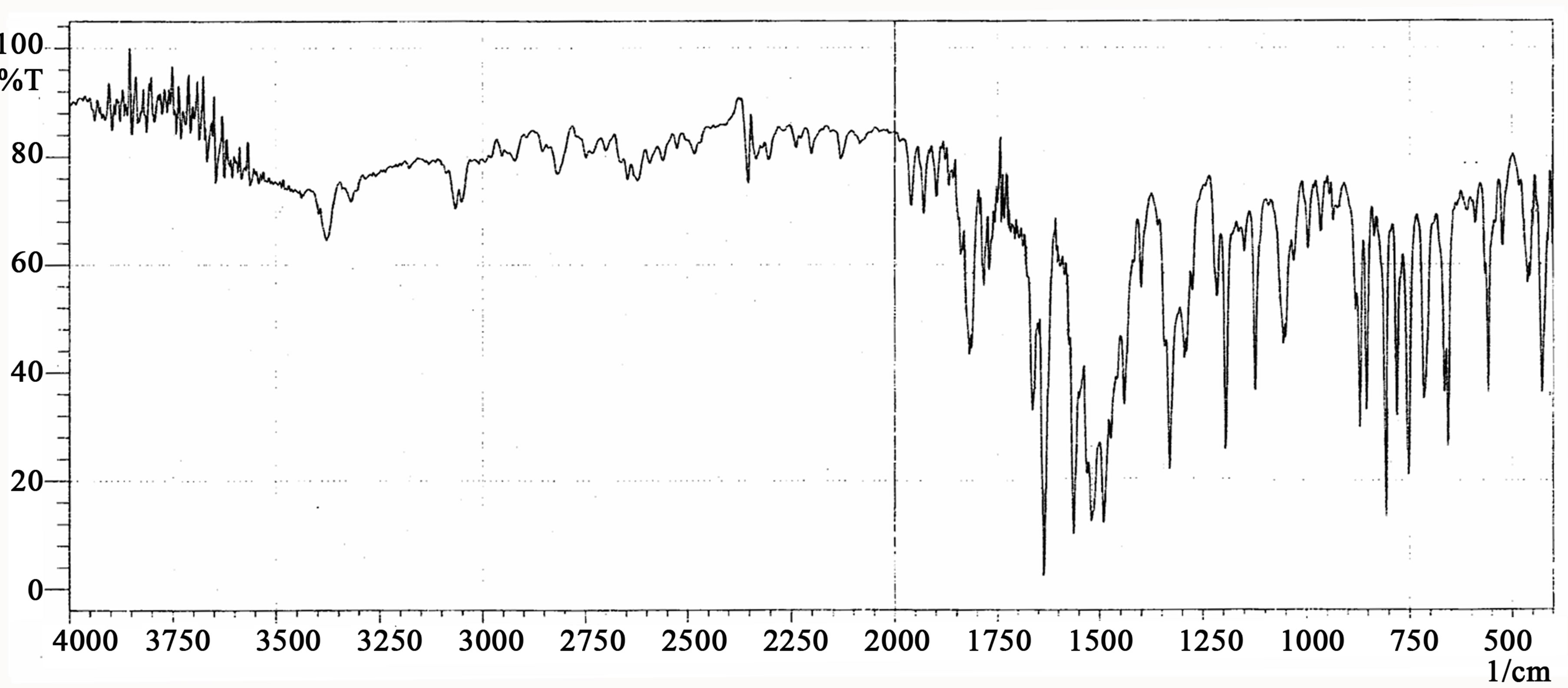
Figure 4. FT-IR spectrum of complex Co(II).

Table 2. FT-IR data of synthesized compounds.
the carboxylato group [24,25]. The IR spectra of the coumarin derivatives shows ≈1612 cm−1 and ≈1745 cm−1 bands corresponding to α, β-unsaturated ketone and lactone carbonyl ketone respectively, on complexation these peaks shifted to a lower frequency ≈1600 cm−1 and ≈1735 cm−1 due to complex formation. In all the complexes, a new band is seen in the ≈ 538 - 546 cm−1 region, which is probably due to the formation of the weak band observed in the ≈ 430 - 455 cm–1 range can be attributed to ν (M-O) [26-27].
3.3. Thermal Studies of Cu(II) Complexes
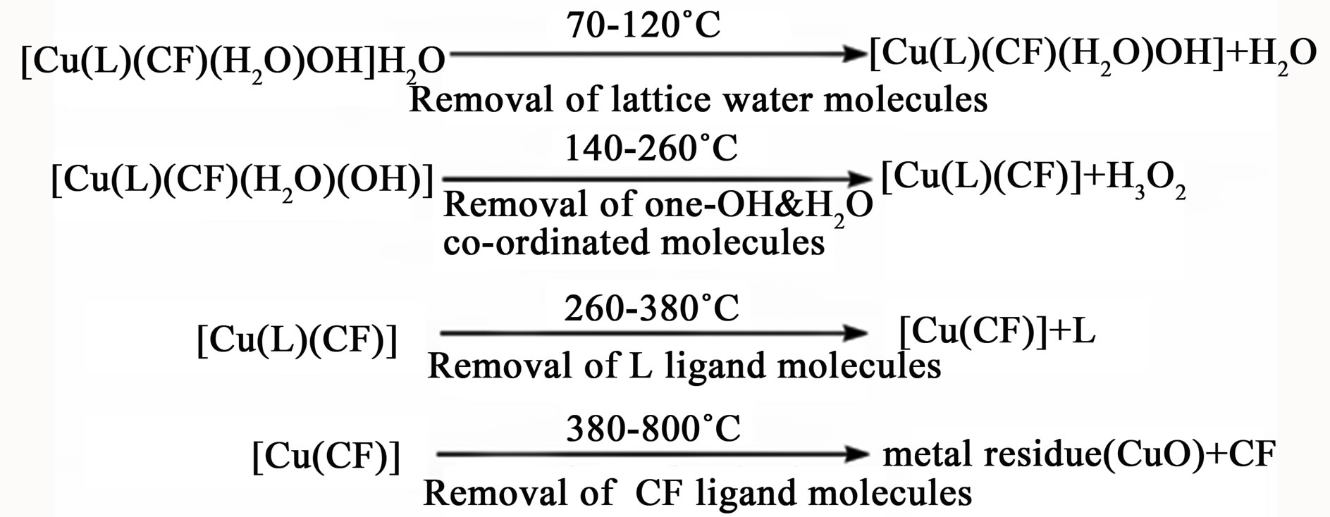
The Thermal behaviour of the complexes was studied using TG whereas TG curves corresponding to the complex (C1) is represented in Figure 5. The thermal decomposition data for all complexes are given in Table 3. The thermal decomposition occurs in four steps in air are observed. According to the mass losses, the following degradation pattern might be proposed for complex [Cu(L)(CF)(H2O)OH]H2O (C1) is represented in Scheme 3. All the compounds decompose with time respectively. Thermal decomposition started by dehydration process and was accompanied by endothermic effect between 80˚C - 110˚C, which was due to loss of one lattice water molecules in first step. The observed mass loss was 2.05% which was nearly equal to theoretical value 2.15%. In the second step, weight loss occur at 220˚C - 250˚C corresponds to loss of one coordinated water and one al of coordinated Ciprofloxacin as well as ligand (coumarins) respectively. As temperature raise, the intermediate complexes [Cu(L)(CF)] (350˚C - 440˚C) and [Cu(CF)] (500˚C - 640˚C) convert to CuO residue of fragments. The observed mass loss for third and fourth stage was 37.68% (calc. 39.60%) and 41.43% (calc. 46.57%) respectively. and remaining weight is in good agreement with copper oxide(CuO).
Scheme 3.Thermal degradation pattern of C1
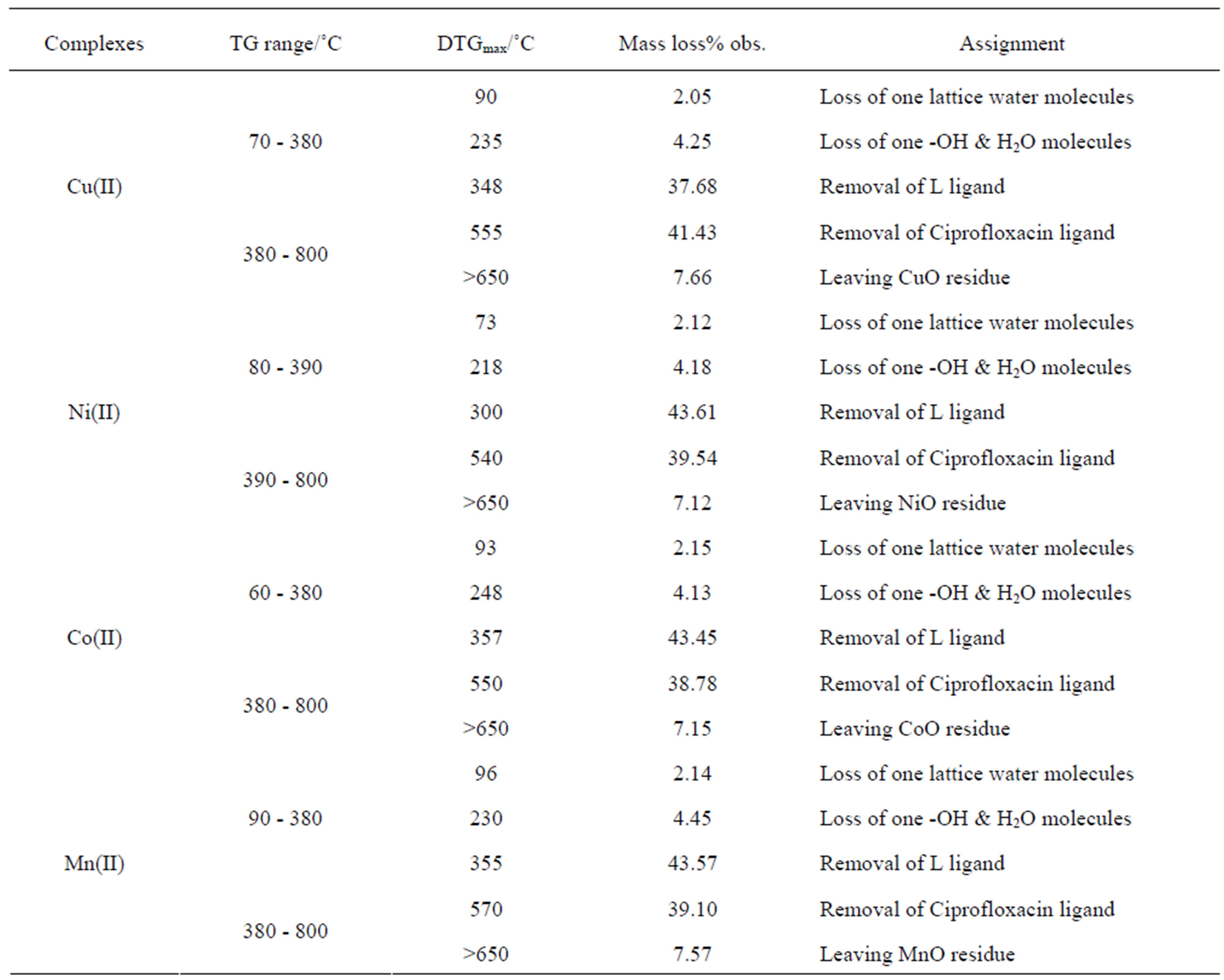
Table 3. Thermoanalytical results (TG and DTG) of metal complexes.
3.4. Electronic Spectra
The Cu(II), Ni(II), Co(II), and Mn(II) complexes show magnetic moments of 1.84. 3.12, 3.88 and 5.92 B.M. respectively which is characteristic of mononuclear, Cu(II) (d9, 1 unpaired electron) octahedral, Ni(II) (d8, 2 unpaired electrons), Co(II) (d7, 3 unpaired electrons), and (d5, 5 unpaired electrons) complexes [28].
The electronic spectral data of the complexes in DMF are shown in Table 4. The Cu(II) complexes display three prominent bands. Low intensity broad band in the region 16,900 - 17,900 cm−1 was assigned as 10 Dq band corresponding to 2Eg→2T2g transition [29]. In addition, there was a high intensity band in the region 22,900 - 27,100 cm−1. This band is due to symmetry forbidden ligand → metal charge transfer transition [30]. The band above 27,100 cm−1 was assigned as ligand band. Therefore distorted octahedral geometry around Cu(II) ion was suggested on the basis of electronic spectra [31]. The electronic spectrum of the Ni(II) complex exhibits three bands at 10,050, 14,925 and 23,529 cm−1, attributable to 3A2g (F) → 3T2g (F) (ν1), 3A2g(F) → 3T1g(F) (ν2) and 3A2g(F) → 3T1g(P) (ν3) transitions, respectively, for an octahedral Ni(II) complex. The electronic spectrum of the Co(II) complex shows two bands at 15,748, 19,230 cm−1 which are assigned to 4T1g → 4A2g(F) (ν2) and 4T1g(F) → 4T1g (P) (ν3) transitions, respectively, as expected for an octahedral Co(II) complex[32,33]. Mn2+ complexes show two bands in the region 18,000 - 20,000 cm−1 and a weak band in the region 23,600 - 24,350 cm−1 for octahedral geometry (Figure 6).

Table 4. Electronic spectral data of the complexes.
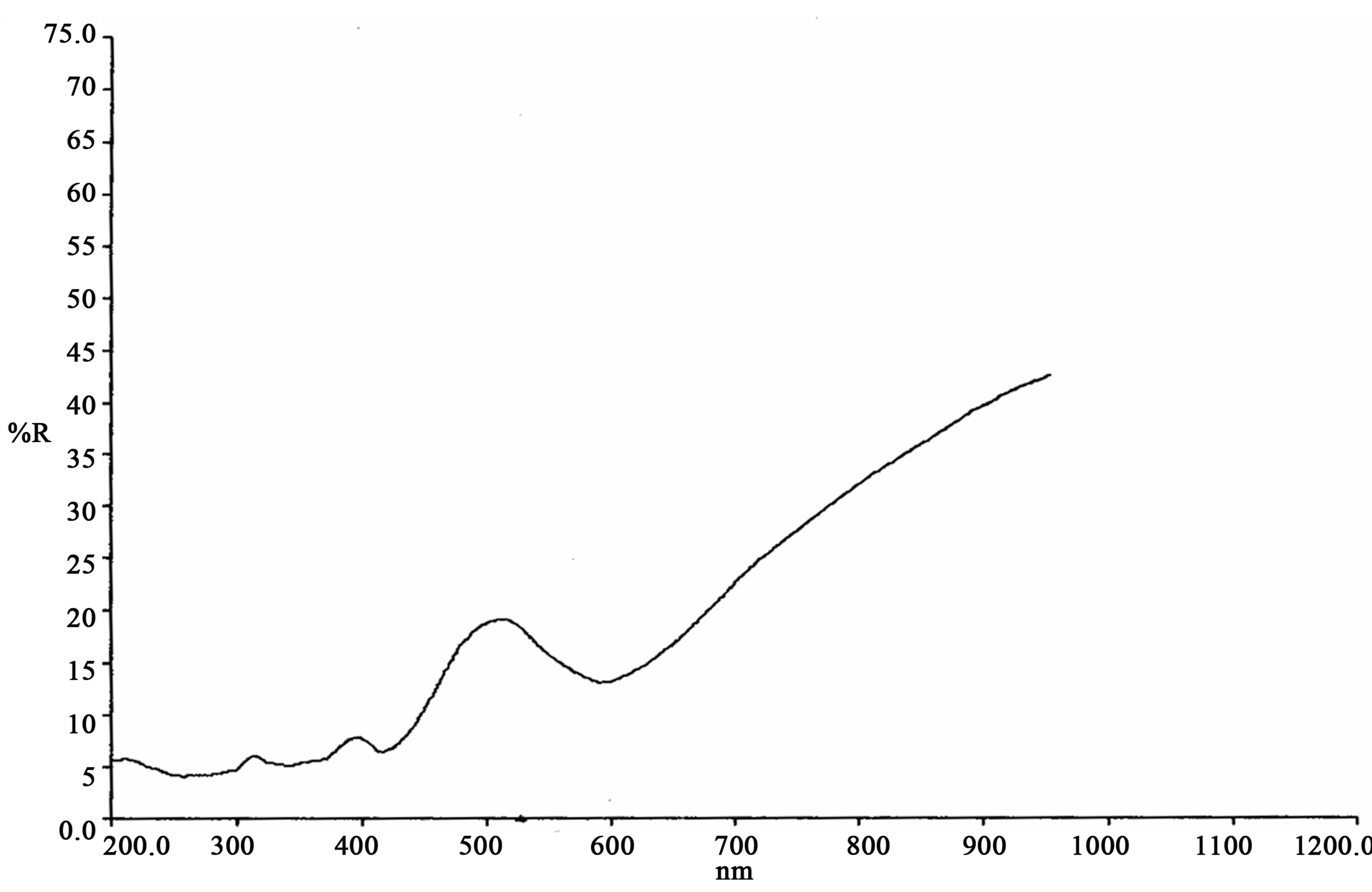
Figure 6. Electronics spectrum of complex Cu(II).
3.5. Antimicrobial Bioassay
The ligand and its metal complexes were screened for their antibacterial and antifungal activities according to the respective literature protocol [34] and the results obtained are p[resented in Table 5. The result was compared with those of the standard drug. All the metal compared with those of the standard drug. All the metal complexes with those of the standard drug. All the metal complexes were more potent bactericides and fungicides than the ligand. Co(II) and Mn(II) complexes were much less bacterial activity than the Cu(II) and Ni(II) complex while Mn(II) complex shows superior antifungal activity compare to other complexes. From Table 5, it can be seen that the highest Antibacterial activity of Cu(II) complex against the bacterium B. subtilis (3.125 µg/ml). On the other hand, Mn(II) complex showed the best activity towards fungi against A. niger (3.125 µg/ml). There was a marked increase in the bacterial and fungi activities of the Cu(II) and Mn(II) complexes respectively, as compared with the free ligand and other complexes under test, which is in agreement with the antifungal and antibacterial properties of a range of Cu(II) and Mn(II) complexes evaluated against several pathogenic fungi and bacteria [35]. For many years it was believed that a trace of Cu(II) destroys themicrobe; however, amore recent mechanism is that activated oxygen in the surface of metal Cu kills the microbe because Cu(II) activity is weak. This enhancement of metal complexes in the activity can be explained on the basis of chelation theory [36]. Chelation reduces the polarity of the metal atom mainly because of partial sharing of its positive charge with the donor groups and possible π electron delocalization within the whole chelate ring. Such a chelation also enhances the lipophililic character of the central metal atom, which subsequently favors its permeation through the lipid layers of cell membrane and the blocking of the metal binding sites on enzymes of microorganism. The variation in the effectiveness of different compound against different organisms depends either on the impermeability of the cell of the microbes or differences in the ribosomes of microbial cells.
3.6. Anti-Tubercular Activity
The Metal(II) complexes were tested for antitubercular activity in order to check the impact of coumarin moiety to with compare activity of Ethambutol (Table 5). The anti-mycobacterial activities of all the synthesized compounds are assessed against M. tuberculosis H37RV at 3.125, 6.25, 12.5, 25, 50 and 100 µg/ml. The Minimum Inhibitory Concentrations of compounds compared with Ethambutol as the standard anti-TB drugs and are summarized in Table 5. Lignad show inhibition at concentration 25 µg/ml. Ni(II) and Co(II) complexes also exhibit activity at same concentration while Cu(II) and Mn(II) complexes have shown enhancement in activity with MIC of 25µg/ml. None of the tested compounds have the inhibition more than standards.
3.7. Antioxidant Studies
A capacity to transfer a single electron i.e. the antioxidant power of all compounds was determined by a FRAP

Table 5. Antimicrobial, anti-tubercular and antioxidant results of compounds.
assay. The FRAP value was expressed as an equivalent of standard antioxidant ascorbic acid (mmol/100g of dried compound). FRAP values indicate that all the compounds have a ferric reducing antioxidant power. The compounds Cu(II) and Ni(II) showed relatively high antioxidant activity while compound Co(II) and Mn(II) shows poor antioxidant power (Table 5).
4. Conclusion
Here Newly the synthesised Cu(II), Ni(II), Co(II) and Mn(II) complexes from biological active Ligand (L) and ciprofloxacin. The structures of the ligand were investigated and confirmed by the elemental analysis, FT-IR, 1H-NMR, 13C-NMR and mass spectral studies. Octahedral geometry were all M(II) complexes assign on the basis of electronic, and TG analysis. All M(II) complexes tested by in vitro antimicrobial, anti-tubercular and antioxidant activity which shows fine results with an enhancement of activity on complexation with metal ions. This enhancement in the activity may be due to increased lipophilicity of the complexes. In review, the antimicrobial testing results reveal that complexes possess higher activity compared to parent ligand.
5. Acknowledgements
We are grateful to The Principal, and management of V. P. & R. P. T. P. Science College, Vallabh Vidyanagar for providing infrastructures and obligatory facilities for research. We are thankful to The Director, SICART, Vallabh Vidyanagar for analytical facilities. Authors are also grateful to The Director, SAIF-RSIC, Panjab University, Chandigarh for NMR analysis. We are articulate our appreciation to IIT-Bombay for TG and DTG scan analysis facility.