1. Introduction
Skin tags, which are also referred to as achrochordons or fibroepithelial polyps (FEPs), are small, soft, fleshcolored to dark brown, usually pedunculated outgrowth of epidermal and dermal tissue that commonly occur on the neck, in the axillae, on the eyelids and in rare cases around genital region [1] [2] .
Diagnosis is made mainly clinically. In most instances, they don’t need to be submitted for microscopic evaluation [3] . However, histological features such as normal or hyperplastic epidermis overlying a fibrovascular tissue core may help for confirmation, especially in unusual cases, that their clinical features may overlap with those of malignant neoplasms [4] .
We present a case of large achrochordon occuring on the labium majus for its unusual site of presentation.
2. Case Report
A 38-year-old, para 5 woman presented with an asymptomatic lesion on her vulva of 3 years duration. The pedinculated lump had increased in size during the 3 years. She had no significant medical or family history. Her menstruation cycles were regular and she had used no medications. She had one vaginal delivery and this childbirth was 14 years ago. According to her history, she had no past history of herpes genitalis or genital warts. On her physical examination, no skin tag anywhere else over the body was found. Pelvic examination revealed an 11 × 5 × 3 cm, brown, pedunculated growth, resembling a scrotal swelling on her left labium majus (Figure 1).
There was no communication with urethra and vagina. There was no inguinal lymphadenopathy. Laboratory results showed a minimally elevated levels of triglyceride. Serological tests for syphilis, hepatitis B and C viruses, HIV 1 and 2 were negative. The lesion was removed under general anesthesia. Histopathology of the specimen revealed polypoidal structure covered by stratified squamous epithelium (Figure 2).
Fibrous stroma and blood vessels in the subepithelium were seen. The diagnosis of achrochordon was confirmed. Written informed consent was obtained from the patient for publication of this case report.
3. Discussion
An acrochordon is a benign tumor that commonly occurs in the natural folds of the skin, such as the neck, axillaes, groin, eyelids and perianal areas [2] . Skin tags are harmless and typically painless. They are not expected to change in size over time [5] . However, our patient described a growth during the 3 years. Even, skin tags are usually small papules or filiform lesions of approximately 2 mm in width and 2 - 5 mm in length, large pedunculated achrochordons can occur rarely, on the lower part of the trunk in particular [6] [7] .
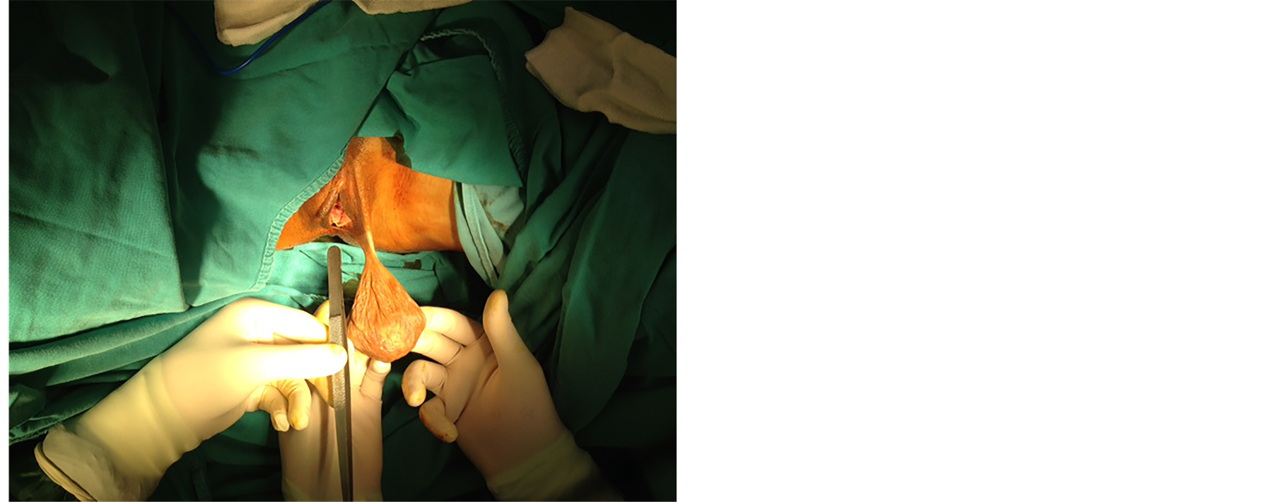
Figure 1. Large polypoidal growth measuring 11 × 5 × 3 cm arising from the left labia majora, resembling a scrotal swelling.
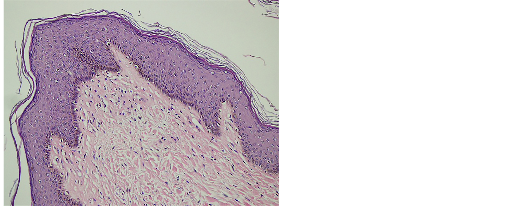
Figure 2. Squamous epithelium covering fibrocollagenous stroma. Hematoxylin and eosin, magnification, 200×.
The etiology of achrochordon is not clearly understood. The main pathologic abnormalities seen in achrochordon are fibroblast proliferation and epidermal hyperplasia. Also, possible involvement of mast cells was reported. It has been proposed that mast cells can induce skin tags through interaction with fibroblasts and keratinocytes [8] [9] . Skin tags have been reported in association with aging, obesity, diabetes mellitus, impaired carbohydrate or lipid metabolism, liver enzyme abnormalities, hypertension, acromegaly, Crohn’s disease, colonic polyps [10] -[12] . Rarely, they can be associated with polycystic ovary syndrome (PCOS) [5] . It is suggested that the origin is most probably from a regressing nevus [13] . In our patient, laboratory tests including lipid profile, fasting glucose, liver function tests were performed and results were within normal limits, but only minimally elevated levels of triglyceride. Her body mass index was 19 and she didn’t meet any diagnostic criteria of PCOS. Because she had no gastrointestinal symptom, Crohn’s disease or colonic polyps were excluded.
Overall, malignant and infectious masses should be considered in the differential diagnosis particularly in genitourinary region, such as squamous cell and basal cell carcinoma, neurofibroma, premalignant fibroepithelial tumor (Pinkus tumor), genital and non-genital warts [14] [15] . Also, a skin tag may be a presenting sign of nevoid basal cell carcinoma syndrome or squamos cell carcinoma can arise from a skin tag [1] [16] . However, nevoid basal cell carcinoma syndrome is seen in childhood and the tumors associated with the syndorme are usually small, unlike our case.
Premalignant fibroepithelial tumor (Pinkus tumor), considered to be a very rare variant of basal cell carcinoma and may resemble an acrochordon while it is a benign-appearing, pink, pedinculated tumor [17] . Even though, these tumors are usually localized on brachium, gluteal region, mesogastrium, and the abdomen, a case of giant Pinkus tumor on the genital region was presented [18] .
Neurofibromas may be found at any side including vulva. Usually these tumors don’t exceed 4 cm, however, a giant neurofibroma, alike a skin tag, was reported by Amita M. et al. [19] . These tumors can grow rapidly and diagnosis is extremely difficult. Histological examination is must [19] .
Despite malignancies associated with acrochordon as described above, the general population has an incidence of skin tags of 46% and malignancy is rare in skin tags with an incidence of 5/1335 [3] [20] . Because of low probability of malignacy, these lesions can be left untreated in most patients. However, it is obvious that the lesion of such size requires excision, even when it is asymptomatic. Because our patient described a growth during the last 3 years and the tumor was in genitourinary region, malignancy was kept in mind and surgical excision was performed.
4. Conclusion
In conclusion, we present a rare case of large achrocordon of labium majus, which can be confused with a malignancy. Pathological examination of vulvar skin tags may be necessary because of its wide range, so excision of the tumor is recommended.
NOTES
*Corresponding author.