Comparison of Magnetic Resonance Imaging and Arthroscopy Findings in the Diagnosis of Meniscus and Anterior Cruciate Ligament Injuries ()

1. Introduction
The knee joint injuries are one of the most common injuries of the human body. They mostly occur during sport activities in younger population. The knee joint, itself, is not protected by any muscular thickening in frontal area and it has a tendency toward injuries due to this lack of reinforcement. Meniscus and ligaments tearing result in the twisting injuries [1] . In 55% - 65% of the cases injuries of the meniscus are often combined with anterior cruciate ligament (ACL) injuries. Since the medical examination is often insufficient to make a clinical diagnosis, we use additional diagnostic methods such as magnetic resonance imaging (MRI) and arthroscopy [2] . MRI is noninvasive diagnostic method, which has an important role in detecting knee injuries [3] . We use MRI not only to make a diagnosis, but to decide if a patient needs further diagnostic methods, such as arthroscopy [4] . Arthroscopy is also used as a treatment method, but it has a disadvantage of being an invasive method [1] . Arthroscopic examination lets us evaluate, visualize, and confirm if the diagnosis based on clinical examination and MRI is correct [4] . Typical symptoms of the twisting knee injuries are pain and blockade. The tear can be longitudinal pericapsular, radial, parrot beak or, bucket handle type. The tear happens usually to younger athletes [5] . In relation to lateral meniscus (LM) injuries, medial meniscus (MM) injuries are much more common. A strong torsional force on the meniscus occurs during sudden abduction and external rotation of the lower leg when the lower leg is in semi-flexion and fixed with the foot [6] . LM injuries occur because of a varus and internal rotation, usually due to a fall on the bent leg; are less frequent and commonly presented as longitudinal tears [7] . ACL injury occurs by an indirect mechanism in case of twisting, flexion, and in cases of contact and deceleration. There is the pain feeling that something is broken (“cracked”) in the knee, functional disability of the knee and acute painful knee effusion in the next few hours, up to 24 hours [8] .
The aim of this work is to compare magnetic MRI and arthroscopic examination in meniscal and ACL injuries.
2. Materials and Methods
This retrospective study was conducted at the Clinic of Orthopedic Surgery and Traumatology of the University Clinical Center of Vojvodina in Novi Sad, during the period from September 1, 2022 to December 31, 2022. The study included 100 patients. As materials, we used knee arthroscopy findings with previously performed MRI findings. Only patients with knee injuries were included in the study. Exclusion criteria were: previous knee surgeries, absence of MRI findings and presence of degenerative changes in the knee. All MRIs were interpreted by the same radiologist and weren’t older than 3 months. All operations were performed under regional anesthesia by the same surgical team. When it comes to methods, statistical data processing was performed in the IBM SPSS Statistics program using the Ks2 test as well as the Wilcocon signed-rank test.
3. Results
This study involved 100 patients, of whom 63 were male and 37 female. The average age of the patients was 32 years (20-52). An analysis of the MRI findings in 28 subjects revealed an ACL injury, which was also confirmed by the arthroscopic findings (Table 1, Chart 1). In 33 patients, in addition to the ACL injury, the MRI findings also indicated a meniscal injury, a medial one in 18 of them, and lateral one in 14 patients. In one patient, in addition to the ACL injury, the MRI findings also verified injury in both menisci. The same was verified in the arthroscopic findings, with the exception of two subjects in whom the lesion of the lateral meniscus was not observed arthroscopically, but was described in the MRI findings (Table 1, Chart 2). An isolated meniscal lesion was observed in 39 patients, 20 in the medial meniscus and 17 in the lateral meniscus (Chart 3). Injury of both menisci was observed in two patients. The same was verified by arthroscopic findings, except in 3 patients with an MRI-verified lesion of the medial meniscus.
![]()
Table 1. MRI and arthroscopic findings in patients who were included in this study.
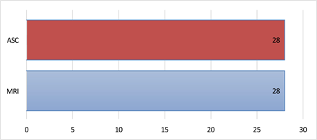
Chart 1. MRI and arthroscopy findings in patients with LCA injury.

Chart 2. MRI and arthroscopy comparison findings in patients with LCA and meniscal injury.

Chart 3. MRI and arthroscopy comparison findings in patients with meniscal injury.
4. Discussion
Often we can not make a diagnosis of meniscus and ACL injury with certainty based on clinical examination alone. To make a definitive diagnosis, we have to use auxiliary diagnostic methods, such as MRI and arthroscopy. Since both methods have some shortcomings, this study was created with the aim of comparing the arthroscopic finding with the MRI finding.
MRI analysis is more comprehensive and provides a more detailed insight into both intraarticular and extra-articular structures of the knee joint. Since arthroscopy shows the intraarticular components of the knee joint, changes in the knee’s soft tissue structures, such as the medial collateral ligament, the structures of the posterolateral angle, and the extensor apparatus are difficult to detect. Also, arthroscopy can’t detect infiltrative changes in the bone marrow, which almost always occur when the meniscus is injured. MRI is a more sophisticated method when it comes to diagnosing changes in the knee synovium [9] .
Arthroscopy provides a dynamic assessment of the soft tissue structures of the knee, while MRI doesn’t. Adequate assessment of osteochondral lesions and meniscal injuries can be achieved with both arthroscopy and MRI. Arthroscopy is a more accurate method when it comes to postoperative monitoring of findings on the menisci. After meniscal surgery, during healing, the signal amplification persists on the MRI findings, which is also noticeable in the case of repeated injuries to the meniscus. Arthroscopy provides a more precise insight into whether it is postoperative healing or re-injury [10] . According to modern studies, the oblique anatomical position in the sagittal plane is one of the reasons for the difficult interpretation of MRI findings in patients with an ACL injury. In our study, all MRI findings were in complete agreement with the arthroscopic findings when it came to ACL injury. Dynamic arthroscopic visualization has greatly increased the diagnostic accuracy of arthroscopy when it comes to ACL injuries. Functional status, ACL injury as well as meniscal injuries can be better demonstrated by arthroscopy although MRI is a very sensitive and specific diagnostic method. The degree of medial and lateral joint gap during arthroscopy serves as an indicator for the injury severity to the collateral ligaments of the knee. During arthroscopy, when the knee is bent at 900, the functionality of the posterior cruciate ligament (PCL) is assessed by performing the posterior drawer test [11] .
The knee is a complex joint, therefore its anatomy is also complex. There are many structures within the knee joint that can not be shown with either arthroscopy or MRI. For example, the posterior horn of the medial meniscus, which plays an important role in limiting anterior translation of the tibia, can sometimes be difficult to detect from standard anterior arthroscopic views.
In this study, the number of diagnosed injuries was approximately the same with both diagnostic methods, which indicates their equal diagnostic value. The results obtained by this study are in accordance with other studies conducted so far on this topic [12] [13] [14] . Using the Wilcoxon signed rank test in the correlation of MRI and arthroscopic findings, a slightly higher p value was obtained for the medial meniscus compared to the lateral meniscus as well as the ACL. Duong et al. [15] reported similar results in their retrospective study.
The analysis of both diagnostic methods in our study recorded a greater number of medial meniscus injuries, which is in accordance with literature data [12] [13] . In the study by Mirković et al. [14] , a higher number of lateral meniscus injuries were recorded, which they attributed to a small sample of patients.
The disadvantage of this study is a small sample of patients. As a disadvantage, it could also be considered that all MRI examinations were performed using the same technique.
5. Conclusions
When it comes to only isolated LCA injuries, the MRI findings fully correspond to the arthroscopic findings.
If, along with the LCA injury, there is also a meniscal injury, or, there is only a meniscal injury, the MRI findings almost completely correlate with the arthroscopic findings.
From all of the above, we can conclude that MRI and arthroscopy are equally valid diagnostic methods for making a definitive diagnosis in a patient with an ACL injury and meniscal lesions.