Fournier’s Gangrene in a Child Hospitalised in the Paediatric Emergency Department of the Gabriel Touré Teaching Hospital ()
1. Introduction
Necrotizing fasciitis is a severe soft tissue infection involving the superficial and deep fascia. Fournier’s Gangrene is a form of genital, perineal and perianal necrotising fasciitis that results from a polymicrobial infection whose source may be genitourinary, colorectal, cutaneous or idiopathic [1] . Despite its relative rarity, Fournier’s gangrene was and remains a formidable disease with severe complications and a high mortality rate [2] [3] . Although Fournier’s gangrene was first described by Baurienne in 1764, it was the French venereologist, Jean Alfred Fournier, who provided a detailed description of the disease in 1883 as fulminant gangrene of the penis and scrotum [4] . Predisposing factors for Fournier’s gangrene include abscesses, omphalitis and diaper rash, surgical procedures such as circumcision, burns, insect stings, anorectal trauma, and nephritic syndrome [5] [6] . Diagnosis is based on clinical assessment. The classic signs are pain, swelling and erythema of the perineum and scrotum. There may be a foul-smelling, dishwasher-like discharge and crepitus may be felt on examination in 19% - 64% of patients. Systemic septic shock can lead to multi-organ failure [7] . Management of Fournier’s gangrene includes early and aggressive resuscitation with fluids, broad-spectrum intravenous antibiotics and surgical debridement of necrotic tissue [8] .
The objective was to report a clinical case of Fournier’s gangrene in an infant following an insect bite.
2. Patient and Observation
This is a 12-month-old boy with no history of surgery, injury to the perineum or lower abdomen, or catheterisation. It is correctly vaccinated according to the extended vaccination programme in force in our country, including the tetanus vaccine at the 6th, 10th and 14th week of life. He was brought by his parents to the paediatric emergency room of the Gabriel Touré University Hospital for an extensive ulcero-necrotic wound and persistent fever, which had been evolving for more than two weeks. The history of the parents (who are farmers and live in a rural area in precarious socioeconomic conditions) reveals a notion of being bitten by a scolopendra (Picture 1) in the right inguinal fold for three weeks.
[The centipede is a carnivorous and venomous arthropod of the family Myriapoda. Its long and very narrow body is made up of twenty-one rings each bearing a pair of legs. The centipede makes two simultaneous movements during its attack, a pinch and a bite. The person found with two small incisions at the level of the wound. The venom induces a release of histamine not mediated by antibodies. The signs are: pain, a local inflammatory reaction (erythema and edema).]
The appearance of a painful swelling at the puncture site plus fever prompted the parents to initiate a traditional treatment based on ointment (composition unknown) and massage. The disastrous evolution of the swelling with the development of necrosis was the reason for the hospital consultation.
The physical examination on admission shows:
A poor general condition, weight at 7 kg 400, height at 76 cm (weight/height ratio < −3 ZScore, reflecting severe acute malnutrition), temperature at 38.8˚C, heart rate at 96 beats/min, respiratory rate at 36 cycles/min and oxygen saturation at 98% under room air.

Picture 1. The centriped.
Moderate pallor of the conjunctivae, deep right inguinal ulceration with tissue necrosis of the dermis and subcutaneous tissue in multiple foci reaching the scrotum with externalization of the right testis and extending to the abdominal wall.
A peri-necrotic palpation that finds crepitus and discharge from the wound, emitting a foul odour (Picture 2).
The diagnosis of Fournier’s Gangrene on a site of Severe Acute Malnutrition (SAM) was retained. Some complementary examinations (entirely at the expense of the parents) were carried out, the results of which are as follows:
· a blood glucose level of 5.05 mmol/l;
· a NFS objective hyperleukocytosis at 22,400/mm3 (normal value: 4000 to 10,000), predominantly granulocytic (40.6%), normochromic, normocytic anaemia with a haemoglobin level at 7.1 g/dl, platelets at 195,000/mm3;
· blood cultures returned sterile;
· negative HIV serology.
Probabilistic antibiotic therapy was instituted (Ceftriaxone and Metronidazole) in addition to packed red blood cell transfusion and nutritional recovery with F75 milk.
The child was then taken successively to the urology department of the CHU Gabriel Touré and to the dermatology hospital for debridement and grafting (Picture 3 and Picture 4), during which pressure on the peri-ulcer skin caused frank yellow pus to flow, which was taken for cytobacteriological examination and antibiotic susceptibility testing.
Staphylococcus aureus and Bacteroides fragilis were isolated from the specimen. Antibiotic therapy was adapted according to the susceptibility ratio on susceptibility testing and monitored healing (wound healing, grafting, reconstructive surgery) was completed after 104 days of evolution.
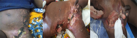
Picture 2. Aspects of gangrene before debridement.
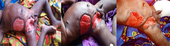
Picture 3. Aspects of gangrene after debridement.
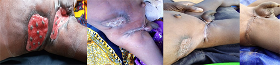
Picture 4. Different stages of wound healing.
3. Discussion
Fournier’s gangrene is a severe and progressive form of necrotising fasciitis, affecting the external genitalia, perineal or perianal regions [3] . It is usually a polymicrobial infection caused by the synergistic action of aerobic and anaerobic organisms [8] [9] . The organisms usually incriminated are: E. coli, Bacteroides, Staphylococci, Streptococci, and Clostridia [5] . This suspicion was verified in our case with the detection of Staphylococcus aureus and Bacteroides fragilis in the discharge from the lesion. Both sexes and all ages can be affected [4] . However, it is relatively rare in children, the majority of cases reported in the literature being in infants under 3 months of age [5] [6] . Its sometimes atypical presentation often leads to a delay in diagnosis, which can be accompanied by a high case fatality of up to 90% due to the development of septic shock and its associated complications [1] [2] . Most studies report mortality rates between 20 and 40%, with a range of 4% - 88% even in developed countries [4] [10] . Colorectal (30% - 50%), urogenital (20% - 40%), skin infections (20%) and local trauma are frequently identified as causes of Fournier’s gangrene. In children, it is most often related to trauma, insect bites as in our case, circumcision, burns and systemic infection [4] [11] . The diagnosis of Fournier’s gangrene is primarily clinical, and in most cases imaging is neither necessary nor desirable [4] . Its clinical presentation is variable but often the diagnosis is made with oedema, erythema, pain, fever and crepitus which is a common feature, present in 50% - 62% of cases due to the presence of gas-producing organisms [7] [12] . The combination of aerobic and anaerobic bacteria can lead to the production of several enzymes, such as collagenases and heparinases, which can result in tissue destruction and rapid progression of the infection [10] [13] . The infection can thus involve the scrotum, the penis and may extend to the anterior abdominal wall as is evident in our case [13] [14] . Involvement of the testis is rare as it has an independent blood supply to the affected area [8] . Empirical broad-spectrum antibiotic therapy should be started without delay and should target gram-negative, gram-positive and anaerobic organisms [7] . Surgical debridement of necrotic tissue controls the spread of infection and reduces systemic toxicity [5] [15] . Several cases of Fournier’s gangrene in infants have been successfully treated with surgical debridement and parenteral antibiotics [5] . However, modern reconstructive techniques, such as skin grafts and flaps, can achieve reliable coverage of large tissue defects and acceptable cosmetic results [9] [13] . In our case, the initial early antibiotic therapy was a combination of Ceftriaxone + Metronidazole, which we subsequently adapted to the susceptibility test according to the sensitivity ratio of the isolated germs. The patient received nutritional support according to the malnutrition management programme in force in our hospital. If not treated aggressively, the mortality associated with Fournier’s gangrene is high, requiring multidisciplinary management [9] [12] . Thus, for our patient, surgical debridement was performed in a urology department and skin grafting in a dermatology hospital, resulting in near perfect healing in 104 days.
4. Conclusion
A poorly managed insect bite can be a cause of Fournier’s gangrene in a malnourished setting. However, rapid diagnosis, early antibiotic therapy and multidisciplinary wound management were key to our success.
Ethical Considerations
We required the informed consent of the parents for the writing of this manuscript and the use of the images.