
1. Introduction
Teratoma is a rare tumor of the pericardium or heart, usually affecting children under one year old. Most often, the teratoma is localized in the pericardium, attached to the root of the aorta or the pulmonary artery, and it is accompanied by usually abundant pericardial effusion. It is a large germ cell tumor, the size of which generally varies from 2 to 9 cm, but can go up to 15 cm [1]. Histologically, the tumor is multicyclic and multilobed. Microscopic analysis finds an association of tissues derived from the 3 embryonic lines [1]. Recurrence after surgical removal, even incomplete, is exceptional [1] [2].
Despite the rarity of this tumor, we must think about it.
2. Observation
It was a newborn male, 3 weeks old, and the third in a family of three children, the first two of whom were alive and well. There was no notion of consanguinity between the parents. The pregnancy was followed with 4 prenatal consultations, there were no particularities on the assessments carried out. Obstetric ultrasound was not performed. The delivery took place at term vaginally on a vertex presentation without any incident. The newborn was eutrophic and extra-uterine adaptation was satisfactory with an Apgar score of 8/10 at the 1st minute then 10/10 at the 5th minute. He was admitted to our unit at 3 weeks of age for progressively worsening respiratory distress, refusal to suckle, incessant crying. The examination revealed a temperature of 36.8˚ Celsius, a heart rate of 168 beats/minute and a respiratory rate of 79 cycles/minute. Weight was 3500 g, height 50 cm and head circumference 33 cm. Physical examination revealed mucocutaneous pallor, severe respiratory distress with signs of struggle, muffled heart sounds, hepatomegaly with hepatojugular reflux. The femoral pulses were malperceived and the pulmonary fields were free. The biological examinations showed a slight hyperleukocytosis with neutrophilic predominance, anemia with a hemoglobin level at 11.7 g/l normochromic, normocytic, a CRP at 13 mg/l. He benefited from the frontal chest X-ray showing global cardiomegaly with a cardio-thoracic index of 0.92 (Figure 1). Two-dimensional echocardiography
![]()
Figure 1. Frontal chest X-ray: cardiomegaly with an ICT of 0.92.
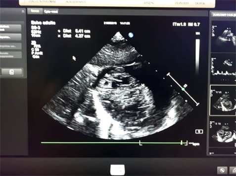
revealed an intrapericardial mass (54.1 mm × 42.7 mm) (Figure 2), a very abundant pericardial effusion compressing the heart, with a heart without anatomical malformation and normal contractility. Computed tomography showed a multicystic intrapericardial mass with a capsule, adhering to the wall of the ascending aorta and compressing the atrium and the right ventricle (Figure 3). The alpha feoprotein assay was normal.
The ultrasound-guided pericardial puncture had brought back about 120 ml of serofibrous fluid.
The newborn was referred to the thoracic and cardiovascular surgery department where he underwent complete resection of the mass (Figure 4). Histology confirmed the diagnosis of teratoma, but histopathology images are not available.
![]()
Figure 2. Cardiac ultrasound: intrapericardial mass compressing the right cavities and abundant pericardial effusion.
![]()
Figure 3. Chest CT scan: cross-sections showing mass and effusion.
![]()
![]()
Figure 4. Complete resection of the mass.
3. Discussion
Cardiac tumors are rare in the pediatric population. In a series of 11,000 autopsies, the incidence was estimated at 0.027%. These tumors are most often benign and primitive [1] [3]. Unlike adults, metastatic cardiac locations are exceptional. The most common histological type is rhabdomyoma, followed by fibroma and teratoma. In the fetus, the teratoma ranks second in frequency behind the rhabdomyoma. Teratomas correspond to 19% to 25% of these [3]. Intrapericardial teratomas are rare lesions representing approximately 10% of all mediastinal masses in children. They were first described by Joel et al. in 1890 [4]. Most often, the teratoma is localized in the pericardium, attached to the root of the aorta or pulmonary artery. It is then accompanied by a generally abundant pericardial effusion. In fetal life, effusion or mass effect can cause extrinsic compression on the heart and tamponade [5]. Much less frequently, the teratoma can be intracardiac, appended to the atrial or ventricular wall [1].
The advent of obstetric ultrasound over the past two decades has enabled prenatal diagnosis and early treatment. Several cases of intrapericardial teratomas diagnosed antenatally have been reported in the literature since 1983 [5] [6] [7].
They can be the cause of non-immunological fetal anasarca, which, if it occurs early and is not treated in utero, can lead to fetal death by cardiac compression or tamponade [8].
The most frequently found prenatal therapy for symptomatic forms (anasarca and suspicion of cardiac tamponade) remains pericardiocentesis, which can be single or repeated [8] [9]. In utero laser treatment has been reported by Liddle et al. [10] at 25 WA by ultrasound-guided laser destruction of the feeding vessels and elective perforation of the cystic parts of the tumor, with birth by caesarean section at 35 WA and six days of a healthy child to date.
The antenatal diagnosis which would have allowed us to anticipate the care from birth before the appearance of complications could not be done in our patient. Obstetric ultrasound was not performed.
In newborns, the symptoms are generally severe with clinical signs of tamponade: respiratory distress, cyanosis and heart failure. Cases of sudden perinatal death have been reported [1].
Vladimir Milovanovic et al. [11] reported a similar neonatal case at delivery, a 2170 g female newborn with diffuse edema in severe cardiorespiratory distress and required resuscitation and intubation.
On the other hand, in our patient the clinical manifestations of cardiac dysfunction were late, during the third week of life. Amadou Gabriel Ciss et al. [2]. A 15-day-old male newborn weighing 3.5 kg was admitted with signs of heart failure and tamponade.
Ultrasound shows the large inhomogeneous multicystic intrapericardial tumour, hanging from one of the two large arterial trunks that it can eventually displace or compress, and usually abundant pericardial effusion, sometimes causing signs of tamponade [1]. Two-dimensional echocardiography is the main diagnostic imaging modality [12]. In developing countries, explorations including magnetic resonance (MRI) or CTScan are not immediately accessible for the majority of the population. The only alternative for us in this emergency case of cardiac tamponade is pericardial drainage. MRIs and CT scans have advantages in assessing the relationship between large tumors and adjacent tissues [12].
The surgical procedure for removal of an intrapericardial teratoma is usually simple. The mass is rarely associated with intracardiac disease and its removal does not require cardiopulmonary bypass. The mediastinum should be explored to verify the absence of another teratoma [13]. In our patient the mass was completely removed, the fixation of the aortic wall was excised without arterial lesions.
Once resected, the prognosis is generally good, as most tumors are benign and require no further treatment [1] [14].
4. Conclusion
Pericardial teratoma has an often-severe clinical expression in neonates that can lead to death by adiostole, but the prognosis is favorable after resection of the mass. Prenatal diagnosis is possible and allows early treatment.
Consent
The examination of the patient was conducted according to the principles of the Declaration of Helsinki.
The authors certify that they have obtained all appropriate patient consent forms, in which the patients gave their consent for images and other clinical information to be included in the journal. The patients understand that their names and initials will not be published and due effort will be made to conceal their identity, but that anonymity cannot be guaranteed.