Synthesis, Characterization, Antibacterial, and Antifungal Activities of Cobalt(II), Nickel(II) and Copper(II) Complexes with 3-thioacetyl-2-amino-1,4-naphthoquinone and 2-benzoyl-3-amino-1,4-naphthoquinone Ligands ()
1. Introduction
Naphthoquinones are widely distributed in plants, fungi, and some animals. Their biological activities have been studied including their effects on prokaryotic and eukaryotic cells, [1]. Naphthoquinones are naturally occurring in plant origin that has antibacterial effects on several species of both aerobic and anaerobic organisms, and toxins derived from naphthazarin [2]. (5,8-dihydroxy-1,4- naphthoquinone) are produced by attack plants, other fungi and bacteria [3]. The antimicrobial activity of the natural naphthoquinone products alkannin and their derivatives has been investigated, in general, they are active against Gram-positive bacteria such as Staphylococcus aureus, Enterococcus faecium, and Baciilus subtilis, but they are inactive against Gram-negative bacteria [4].
Antibacterial activity has been described for isoxazolylnaphtho-quinones and some hydroxyquinones and their metal complexes [5] [6]. The development of new antimicrobial agents is a research area of the utmost importance. Antimicrobial resistance among key microbial pathogens continues to grow at an alarming rate worldwide. Resistance among strains of S. aureus, Pseudomonas spp, Streptococcus spp and Enterobacteriaceae has been described [7] [8] [9]. The increased prevalence of antibiotic-resistant bacteria emerging from the extensive use of antibiotics may render the current antimicrobial agents insufficient to control at least some bacterial infections. The challenge of synthesizing derivatives of natural antimicrobial naphthoquinones to improve their pharmaceutical properties has been accepted by several laboratories. Indeed, the synthesis and evaluation of antimicrobial activity of bioactive analogs of kigelinone, alkannin, [10] [11], have been reported. In the present study, we describe the antibacterial activity of a new series of 1,4-naphthoquinones. The metal chelates often caused decrease of biological activity. Cobalt complex of (L1 and L2) was the most effective against Gram-positive bacteria, showing MIC values ranging from 375 to 1400 mg/ml. Metal chelates may be useful tools for the understanding of the antimicrobial mechanism of 1,4-naphthoquinones on these bacteria [12].
2. Experimental
2.1. Materials and Reagents
Analytical grade chemical reagents were used. All chemicals used in this study are of the highest purity from commercial suppliers such as Merck; BDH and Aldrich they include 1,4-Naphthoquinone, Thioacetamide and Benzamide, CoCl2∙6H2O, NiCl2∙6H2O and CuCl2∙2H2O. The organic solvents such as an absolute ethanol and methanol, DMF and DMSO are purchased from Alpha Easer.
2.2. Instrumentation
Melting point apparatus (Gallen Kamp, Germany) was used to investigate the melting points. Elemental microanalysis of the separated solid chelates for C, H and N were performed in the Micro analytical Center, Cairo University, using CHNS-932 (LECO) Vario Elemental analyzers. Infrared spectra were recorded on Perkin-Elmer FT-IR type 1650 spectrophotometer in wave number region 4000 - 40 cm−1. The spectra were recorded as KBr pellets. The 1H NMR spectra were recorded using 300 MHz Varian-Oxford Mercury. The deuterated solvent used was dimethylsulphoxide (DMSO-d6) and the spectra extended from 0 to 15 ppm. The electron impact (EI) mass spectra (MS) at 70eV of the tested compounds has been done using MS-5988 GS-MS Hewlett-Packard instrument. TGA was carried out in dynamic nitrogen atmosphere (10 mL∙min−1) with a heating rate of 10˚C min−1 using DTG-50 H Shimadzu simultaneous. The molar conductance of solid chelates in DMF was measured using WPA CM35 Conductivity meter fitted with platinized platinum electrodes. The antibacterial and antifungal activities were evaluated at the Microbiological laboratory, Micro analytical center, Cairo University, Egypt.
2.3. Methods
2.3.1. Synthesis of Free Ligands
1,4-Naphthoquinone (2 g, 0.0126 mol) mixed with an equivalent amount (0.95 g and 0.53 g, 0.0126 mol) of Thioacetamide and Benzamide respectively, then the mixture was added to an aqueous solution of sodium hydroxide and left for two hours on water bath to be fused. The crude product was recrystallized from ethanol and dried under vacuum over P2O5. The yield was 90%. The melting point was measured and listed in Table 1. The procedure cited in respective reference [13] [14].
2.3.2. Preparation of Metal Complexes
The metal complexes were prepared by dissolving (1.088 g and 1.385 g, 0.005
![]()
Table 1. Analytical and physical properties of 1,4-naphthoquinone complexes.
mol) of ligand (L1-L2) respectively in hot ethanol (50 ml) and added drop wisely with stirring to a stoichiometric amount of 1:1 (M:L) molar ratio to (1.189 g, 1.888 g, and 0.852 g, 0.005 mol) of CoCl2∙6H2O, NiCl2∙6H2O and CuCl2∙2H2O respectively. The reaction mixture was refluxed for 40 min and left overnight. The isolated solid complexes were filtered off, washed with distilled water until the solution became colorless and washed with 10 ml hot ethanol-water mixture (1:1) to remove any traces of the unreacted materials. The solid complexes were dried at 70˚C for several hours kept in desiccator containing dry P2O5. Analytical data were listed in Table 1.
2.3.3. Biological Activity
Modified Kirby-Bauer disc diffusion method [15], has been used to determine the antimicrobial activity of the tested samples [16]. Examined 100 μl of the tested bacteria or fungi, it has been found that it was developed in 10 ml of fresh media until they reached a count of approximately 108 cells/ml for bacteria and 105 cells/ml for fungi. 100 μl of microbial suspension was spread onto agar plates corresponding to broth in which they were maintained. Isolated colonies of each organism that might be playing a pathogenic role should be selected from primary agar plates and tested for susceptibility by disc diffusion method of the many media available; NCCLS recommends Mueller-Hinton agar due to it results in good batch-to-batch reproducibility. Disc diffusion method for filamentous fungi tested by using approved standard method (M38-A) developed. For evaluating the susceptibilities of filamentous fungi to antifungal agent, Disc diffusion method for yeast developed by National Committee for Clinical Laboratory Standards using approved standard method (M44-P). Plates inoculated with filamentous fungi as Asprgillus flavus at 25˚C for 48 hours; Gram (+) bacteria as Staphylococcus aureus; Gram(−) bacteria as Escherichia coli, they were incubated at 35˚C - 37˚C for 24 - 28 hours and yeast as Candida albicans incubated at 30˚C for 24 - 28 hours, then the diameters of the inhibition zones were measured in millimeters with slipping calipers of the National Committee for clinical Laboratory Standards [17], have been used standard discs of tetracycline (antibacterial agent), and amphotericin B (antifungal agent) served as positive controls for antimicrobial activity but filter discs impregnated with 10 μl of solvent (distilled water, chloroform, DMSO) were used as a negative control. The agar used is Mueller-Hinton agar that is rigorously tested for composition and pH. Further the depth of the agar in the plate is a factor to be considered in the disc diffusion method. This method is well documented and standard zones of inhibition have been determined for susceptible and resistant values. Blank paper discs (Schleicher and Schuell, Spain) with a diameter of 8.0 mm were impregnated with 10 μl of tested concentration of the stock solutions. When a filter paper disc impregnated with a tested chemical is placed on agar, the chemical will diffuse from the disc into the agar. This diffusion will place the chemical in the agar only around the disc. The solubility of the chemical and its molecular size will determine the size of the area of chemical infiltration around the disc. If an organism is placed on the agar, it will not grow in the area around the disc if it is susceptible to the chemical. This area of no growth around the disc is known as zone of inhibition or clear zone. For the disc diffusion, the zone diameters were measured [18], found that, agar-based methods such test and disc diffusion can be good alternatives because they are simpler and faster than broth-based methods.
3. Results and Discussion
3.1. Elemental Analysis and Physical Properties
The results of elemental analyses and physical properties of the free ligands and its metal chelates shown in Table 1 are in good agreement with those required by proposed formulae. The isolated solid complexes are stable at room temperature, partly soluble in polar organic solvents, but completely soluble in polar aprotic solvents such as DMF and DMSO. Based on the above mentioned results, it can propose the general structural formulae of 1:1 (M:L) complexes (Figure 1, Figure 2).
3.2. Molar Conductivity Measurements
The metal chelates were dissolved in DMF at 25˚C ± 2˚C and the molar conductivities of 5 × 10⁻⁴ M of their solutions were measured by recommended procedure [19]. The obtained molar conductance values are listed in Table 1. The molar conductivity value of Co(II), Ni(II) and Cu(II) chelates of free ligands (L1- L2) were found to be 5.46 up to 9.89 Ω−1∙mol−1∙cm2. The chelates are nonionic in nature and they are considered as non-electrolytes.
3.3. FT-IR Spectroscopy
3.3.1. FT-IR of 3-thioacetyl-2-amino-1,4-naphthoquinone (L1) and Its Metal Chelates
The main FT-IR bands of 3-thioacetyl-2-amino-1,4-naphthoquinone (L1) and its metal chelates are presented in Table 2, Figure 3.
The vibration spectra of ligand Cexhibit a very broad band at 3436 cm−1 assigned to the υ (NH2) stretching vibration of (C2-NH2) of the naphthoquinone. The stretching band at 1563 cm−1 is attributed to υ (C-N) ring stretching vibration. The stretching band at 1259 cm−1 assigned to the υ (C=S) which in good agreement with the structure proposed of the ligand [14]. The FT-IR spectrum of 3-thioacetyl-2-amino-1,4 naphthoquinone (L1) metal chelates exhibits different
![]()
Figure 1. The proposed structure of the prepared metal complexes with 3-thioacetyl-2- amino-1,4-naphthoquinon (L1).
![]()
Figure 2. The proposed structure of the prepared metal complexes with 2-benzoyl-3- amino-1,4-naphthoquinon (L2).
![]()
Table 2. Infrared spectral data of free ligand (L1-L2) and their metal chelates.
bands. A new band appeared at 3547, 3527 and 3538 cm−1 for Co(II), Ni(II) and Cu(II) respectively, that suggests water molecules in the prepared complexes are loosely coordinated or exist as molecules of crystallization. The lower shift of υ (NH2) band in the region at 3400, 3392 and 3351 cm−1 for Co(II), Ni(II) and Cu(II) complexes respectively support the contribution of N-atom of NH2 group in complex formation. The observed stretching vibration band υ (C=S), at 1259 cm−1 of the ligand L1 show lower shift stretching vibration on complexation to Co(II), Ni(II) and Cu(II) at 1229, 1225 and 1227 cm−1 respectively which supports the involvement of Sulphur atom of (C=S) group in the chelation. Also, on complexation the υ (C-N) stretching vibration at 1598, 1576 and 1587 cm−1 for Co(II), Ni(II) and Cu(II) respectively show red shift in these bands, which may be due to increase of bond order of carbon to the nitrogen link following the coordination of the imine nitrogen atom to metal ions. The stretching band of the coordinated water molecules υ (H2O) was observed at 839,824 and 896 cm−1 for Co(II), Ni(II) and Cu(II) respectively. The new bands observed at 590, 589 and 593 cm−1 in all complexes under study may be assigned to υ (M-O) and at 472,470 and 474 cm−1 assigned to υ (M-N).
3.3.2. FT-IR of 2-benzoyl-3-Amino-1,4-naphthoquinone (L2) and Its Metal Chelates
The main FT-IR bands of 2-benzoyl-3-amino-1,4-naphthoquinone (L2) and its metal chelates are presented in Table 2.
The vibration spectra of ligand L2 exhibit a very broad band at 3424 cm−1 assigned to the υ (NH2) stretching vibration of (C2-NH2) of the naphthoquinone. The stretching band at 1663 cm−1 could be assigned to the υ (C=O). The stretching band at 1553 cm−1 may be attributed to υ (C-N) ring stretching vibration. The vibration spectra of the formed complexes of L2, exhibit a broad band around 3542, 3525 and 3538 cm−1 due to the υ (OH) stretching. The lower shift of υ (NH2) band in the region at 3412, 3369 and 3352 cm−1 for Co(II), Ni(II) and Cu(II) complexes respectively support the contribution of N-atom of NH2 group in complex formation. Also, the lower frequency shift of this band confirms our suggestion that NH2 is taking part in coordination. The observed low shift of υ (C=O) 1663 cm−1 band of the ligand L2 on complexation to Co(II), Ni(II) and Cu(II) ions respectively at 1619, 1653 and 1650 cm−1 which supports the involvement of oxygen atom of (C=O) group in the chelation. On the other hand, on complexation the υ (C-N) stretching vibrations at 1570, 1579 and 1580 cm−1 for Co(II), Ni(II) and Cu(II) ions respectively show red shift in these bands, which may be due to increase of bond order of carbon to the nitrogen link following the coordination of the imine nitrogen atom to metal ions [20]. The stretching band of the coordinated water molecules υ (H2O) was observed at 824, 839 and 850 cm−1 for Co(II), Ni(II) and Cu(II) complexes respectively. The new bands observed at 588, 584 and 592 and in all complexes under study may be assigned to υ (M-O) and at 470,480 and 450 cm−1 may be assigned to υ (M-N).
3.4. 1H-NMR Measurements
3.4.1. 1H-NMR of 3-thioacetyl-2-amino-1,4-naphthoqunone (L1) and Its Nickel Complex
1H-NMR spectrum 300 MHz of 3-thioacetyl-2-amino-1,4-naphthoqunone (L1) shows several signals and the resulted data are tabulated in Table 3, Figure 4.
The 1H
NMR
spectrum (L1) shows a singlet signal at δ 2.5 ppm of relative intensity (s, 3H) may be attributed to CH3 protons, a multiplet at δ 7.2 - 7.5 ppm of relative intensity 4H, which may be assigned to four protons (m, 4H) in quinone, Harm. The multiplet signal observed at δ 8 ppm may be assigned to two protons of amino group (m, 2H, NH2) [21]. The comparison of the protons signals,
![]()
Table 3. 1H-NMR data for free ligands (L1-L2) and their metal chelates.
multiplets and the chemical shifts of L1 ligand with its corresponding nickel chelate [NiC12O2H9NS Cl2(H2O)2]∙4H2O is investigated. It has been found that methyl protons of the free ligand are slightly shifted to δ 2.48 ppm. Also, the NH2 protons of the free ligands are slightly shifted to δ 7.89 ppm. This suggests that the metal ion coordination takes place through the nitrogen atom of NH2 group. The proton signal observed at δ 3.29 ppm, which may be assigned to the presence of water molecules is in agreements with the suggested formulae of metal chelates.
3.4.2. 1H-NMR of 2-benzoyl-3-amino-1,4-naphthoqunone (L2) and Its Copper Complex
1H-NMR spectrum 300 MHz of 2-benzoyl-3-amino-1,4-naphthoqunone (L2) shows several signals and the resulted data are tabulated in Table 3.
The 1H
NMR
spectrum (L2) shows a singlet signal at δ 7.29 - 7.51 ppm, which may be assigned to four protons (m, 4H) in quinone, Harm and five protons (m, 5H) in phenyl group. The multiplet signal shown at δ 7.9 ppm may be assigned to two protons of amino group (m, 2H, NH2) [22]. On comparing the investigated 1H-NMR signals of the copper chelate [CuC17H11NO3Cl2(H2O)2] with those of the L2 ligand protons signals, multiplets and the chemical shifts. It has been found that the NH2 protons of the free ligands are slightly shifted to δ 7.82 ppm this suggests that the metal ion coordination takes place through the nitrogen atom of NH2 group. The proton signal observed at δ 3.34 ppm, which may be assigned to the presence of water molecules is in agreements with the suggested formulae of metal chelates.
3.5. Mass Spectroscopic Studies
3.5.1. Mass Spectra of 3-thioacetyl-2-amino-1,4-naphthoqunone (L1) and Its Nickel Complex
The electron impact mass spectrum of [Ni(C12O2H9NS)Cl2(H2O)2]∙4H2O Figure 5 shows many fragment ions which consists of two principle pathways as shown in Scheme 1. The signal that appears at m/z = 468.95 (mole mass = 468.96) may be referred to the appearance of main general molecular weight of metal chelate which undergo two pathways of fragmentation. Pathway I shows a signal at m/z = 324.07 (mole mass = 328.39, RI = 15%); which may be refer to the loss of five molecules of water, CH3 and Cl− ion, followed by the appearance of a signal at m/z = 271.55 (mole mass = 274.93, RI = 15%); which may be refer to the loss of one molecule of water and Cl− ion, followed by the appearance of a signal at m/z = 90.07 (mole mass = 90.75, RI = 34%) as rupture of C11H6NO2 (furo[2,3-b]quinolin-8-olate). Pathway II shows a fragment at m/z = 324.07 (mole mass = 325.99, RI = 15%) may be assigned to the loss of four water molecules, Cl2 followed by the loss of C8H4O2 (benzocyclobutenedione) molecule and two water molecules leaving a fragment give a signal at m/z = 157.57 (mole mass = 157.84, RI = 34%). A signal appeared at m/z = 90.05 (mole mass = 90.75, RI = 34%) as a rupture of C4H5N (pyrrole).
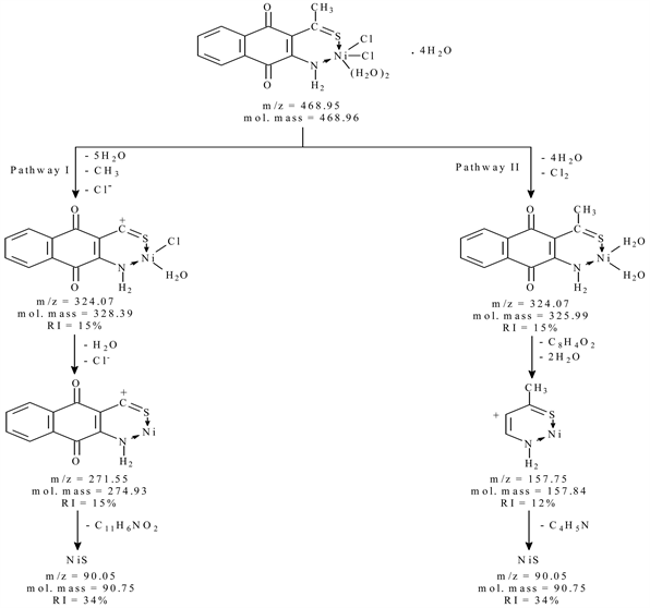
Scheme 1. The mass fragmentation pathways of [Ni L1Cl2 (H2O)2]∙4H2O.
3.5.2. Mass Spectra of 2-benzoyl-3-amino-1,4-naphthoqunone (L2) and Its Cobalt Complex
The electron impact mass spectrum of [CoC17H11NO3Cl2 (H2O)2] Figure 6 shows many fragment ions which consists of two principle pathways as shown in Scheme 2. The signal that appears at m/z = 514.80 (mole mass = 515.20) may be referred to the appearance of main general molecular weight of metal chelate which undergo two pathways of fragmentation. Pathway I stated that; the main metal chelates lose six molecules of water and C6H5 (phenyl radical) leaving a fragment at m/z = 327.07 (mole mass = 330.01, RI = 12%) followed by the appearance of a signal at m/z = 127.89 (mole mass = 126.98, RI = 4%); which may be refer to the loss of C8H4O2 (benzocyclobutenedione) and Cl2. followed by the appearance of a signal at m/z = 76.95 (mole mass = 74.93, RI = 55%) as rupture of C3H2N+ (1-Cyanoacetylenecation). Pathway II shows a fragment at m/z = 311.07 (mole mass = 313.15, RI = 80 %); which may be refer to the loss of three molecules of water, Cl2 and C6H5 (phenyl radical), followed by the appearance of a signal at m/z = 76.95 (mole mass = 74.93, RI = 55%) as rupture of C11H6NO2 (furo[2,3-b]quinolin-8olate) (Figure 5, Figure 6).
3.6. Thermogravimetric Analyses (TGA)
The TGA thermal analyses data of the synthesized metal chelates are tabulated in Table 4.
![]()
Figure 4. 1H-NMR spectrum of [Cu L2Cl2 (H2O)2].
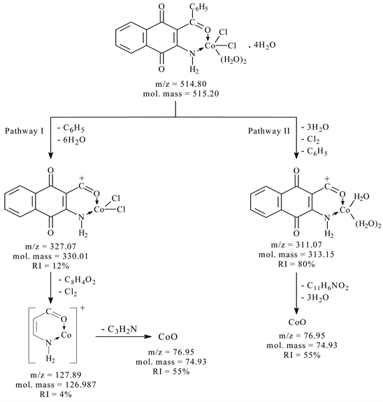
Scheme 2. The mass fragmentation pathways of [Co L2Cl2 (H2O)2]∙4H2O.
![]()
Figure 5. Mass Spectrum of [Ni L1Cl2 (H2O)2]∙4H2O.
![]()
Figure 6. Mass Spectrum of [Co L2Cl2 (H2O)2]∙4H2O.
![]()
Table 4. Thermoanalytical analyses data for newly synthesized chelates of ligands (L1-L2).
3.6.1. Thermal Analysis of 3-thioacetyl-2-amino-1,4-naphthoqunone (L1)-Ni Complex
The thermogram of the complex [NiL1Cl2 (H2O)2]∙4H2O displays four stages of decomposition within the temperature range 50˚C - 980˚C. The first stage between 50˚C - 250˚C corresponds to dehydration process. The experimental mass loss of 16.36% agrees well with the calculated mass loss of 15.36%. The second decomposition stage occurs in the 250˚C - 450˚C range is associated with the burning of the loosely CH3 and two coordinated water molecules. This fact find support from the experimental weight loss value 12.58% and the calculated mass loss 10.88%. The third stage within the temperature range 450˚C - 670˚C corresponding to release of NH2 molecule and Cl2 gas. The further degradation of organic ligand takes place in successive steps within the temperature range 670˚C - 980˚C. The total experimental mass loss is 80.51% agree well with the calculated mass loss of 80.65% and the final product obtained is NiS. All results are shown in Table 4.
3.6.2. Thermal Analysis of 2-benzoyl-3-amino-1,4-naphthoqunone (L2)-Co Complex
The TGA curve for the thermal decomposition process of [CoL2Cl2 (H2O)2]∙4H2O metal chelate illustrates the thermal decomposition occurs through three decomposition steps within temperature range 50˚C - 980˚C. The first step of decomposition occurs within the temperature range 50˚C - 100˚C of estimated mass loss 12.22% (calcd. = 13.98%). This step may be attributed to the separation of four molecules of water of hydration (mole mass = 72.07 g). The second step is followed by loss two coordinated water molecules, C6H5 group and Cl2 gas, within the temperature range 100˚C - 640˚C with mass loss of 35.10% (calcd. = 35.72%), the third step starts at 640˚C and comes to end at 980˚C corresponding to mass loss range 34.32% (calcd. = 35.74%) due to decomposition of the remaining organic ligand molecule. The mass losses are in agreement with calculated mass loss based on the obtained data (Table 4). The final residue is quantitatively proved to be cobalt (II) oxide. The total experimental mass loss 81.64% agrees well with the calculated mass loss of 85.45%.
4. Biological Activity
The comparison of biological activity of the free ligands (L1 up to L2) and its complexes with the standard disc of Ampicillin (antibacterial G+ agent and antibacterial G− agent), Amphotericin B (antifungal agent), towards the different organisms was carried out. The data are listed in Table 5 and shown in Figure 7 & Figure 8). The free ligands and its metal chelates were screened against Staphylococcus aureus and Bacillis subtilis (G+) and Escherichia coli (G−) bacteria and Candida albicans (fungi) to assess their potential antimicrobial agents.
4.1. Biological Activity of 3-thioacetyl-2-amino-1,4-naphthoqunone (L1)
The biological activity of Ligand (3-thioacetyl-2-amino-1,4-naphthoqunone) L1 and its metal complexes shows higher results than that of the free ligand. But all
![]()
Figure 7. Biological activity of (L1) and its metal complexes.
![]()
Figure 8. Biological activity of (L2) and its metal complexes.
![]()
Table 5. Biological activity of L1 and L2 its metal chelates.
of them are lower than standard. Therefore, the biological activity of the complexes follows the order Co(II) > Cu(II) > Ni(II) against Staphylococcus aureus, Bacillus subtilis and Escherichia coli organisms. But with Candida albicans the biological activity follows the order Ni(II) > Co(II) > Cu(II).
4.2. Biological Activity of 2-benzoyl-3-amino-1,4-naphthoqunone (L2)
The biological activity of Ligand (2-benzoyl-3-amino-1,4-naphthoqunone) L2 and its metal complexes shows higher results than that of the free ligand. But all of them are lower than standard. Therefore, the biological activity of the complexes follows the order Co(II) > Cu(II) > Ni(II) against Bacillus subtilis and Escherichia coli organisms for (L2) and complexes Meanwhile, the biological activity of the complexes follow the order Co(II) > Cu(II) > Ni(II) against Staphylococcus aureus. But with Candida albicans the biological activity follows the order Ni(II) > Co(II) > Cu(II).
The importance of this, lies in the fact that these complexes could be applied fairly in the treatment of some common diseases caused by E. coli e.g. Septicemia, Gastroenteritis, Urinary tract infections and hospital acquired infections according to [23]. However, the complexes were specialized in inhibiting Gram-positive and Gram-negative bacterial strains. The importance of this unique property of the investigated complexes lies in the fact that, it could be applied safely in the treatment of infections caused by any of these particular strains. Generally, the activity of the free ligand was increased upon complexation with metal ions; the enhancement in activity can be explained on the basis of chelation theory, reported by [24] [25]. Chelation reduces the polarity of the metal ion considerably, mainly because of the partial sharing of its positive charge with donor groups and the possible p electron delocalization over the whole chelate ring. Chelation not only reduces the polarity of metal ion, but also increases the lipophilic character of the chelate. As a result of this, the interaction between the metal ion and the cell walls is favored, resulting in interference with normal cell processes.
5. Conclusion
In the present study, the free ligands (L1, L2) and its metal complexes Co(II), Ni(II) and Cu(II) respectively were prepared and structurally identified. The structures of free ligands and its metal chelates are proved by elemental analyses and applying spectroscopic measurements (FT-IR, H-NMR, and mass spectra) and confirmed by thermal analyses. The synthesized free ligand is found to be biologically active and their metal complexes showed significantly enhanced antibacterial and antifungal activities against microbial strains in comparison to the free ligand. We are noticed that 1,4-naphthoquinones ligands and its metal chelates are useful tools for the understanding of the antimicrobial mechanism of 1,4-naphthoquinones on these bacteria that will encourage us to continue in this trend.