The Fusability of Erythrocytes as a Method for Evaluating the Instability of Their Membranes ()
1. Introduction
Membrane fusion is ubiquitous cellular process mediating such phenomena as fertilization, exocytosis, phagocytosis, etc. [1]. Fusion can be induced by chemical agents [2]. We have previously shown that La3+ induced extensive fusion of erythrocytes with an altered ATP content [3].
It is known that the lanthanides have an extremely high affinity for phosphatidylserine [4]. It is shown that La3+ at low concentrations induces fusion phosphatidylserine vesicles [5]. Exposure of phosphatidylserine on the surface of lipid-symmetric erythrocytes may be responsible for their enhanced fusion [6]. Phosphatidylserine externalization leads to erythrocyte disintegration, or, in the presence of macrophages, to macrophage ingestion of dying erythrocytes [7,8]. It has been shown that chronic inflammation [9] and heart shock [10] greatly increase the frequency of cell fusion.
When whole blood is stored in a preservative medium, there may be morphological, biochemical, and metabolic change in the red blood cells [11].
Diabetes mellitus is the most prevalent metabolic disease and represents a serious clinical and public health problem. Increased oxidative stress and decreased life span of erythrocytes are reported in patients with type 2 diabetes. A positive correlation between lipoperoxidation and phosphatidylserine externalization in erythrocytes of patients with diabetes was found [12].
In the present study, we investigated the influence of La3+ on the fusion of erythrocytes blood storage and erythrocytes of patients with diabetes.
2. Methods and Materials
This study included 8 healthy volunteers and 5 type 2 patients with diabetes. Blood from all study subjects was obtained by venous puncture in vacuum tubes containing 3.2% sodium citrate (in а ratio 9:1) as an anticoagulant. Each sample was centrifuged (3000 × g, 20 min), and the
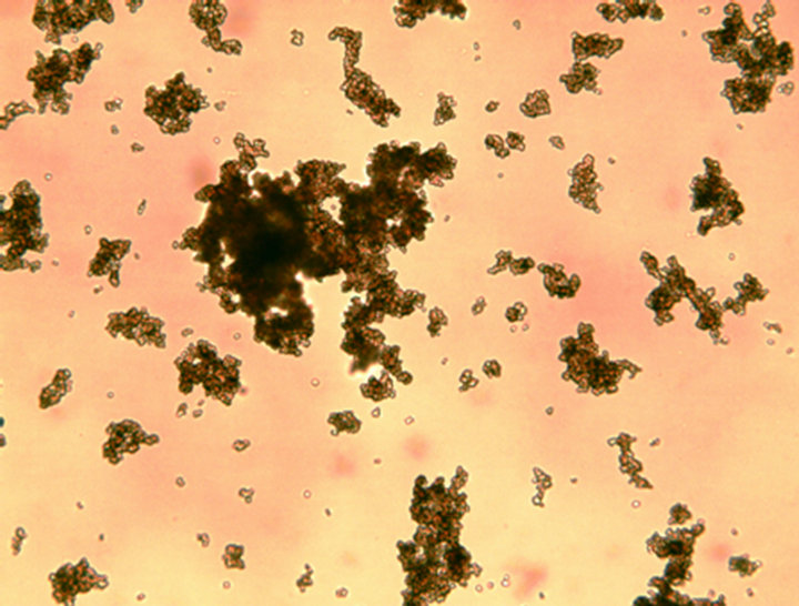
Figure 1. Influence of La3+ (150 µM) on fusion of normal of human erythrocytes. Incubation for 120 min at 37˚C × 100.
plasma and white cells were carefully removed by aspiration to avoid loss of erythrocytes. Erythrocytes were washed three times with a physiological solution (150 mM). Then 0.05 ml of washed red blood cells was resuspended in 10 ml of 10 mM Tris-HCl (pH 7.4) containing 150 mM NaCl. To 0.9 ml of the erythrocyte suspension in plastic tubes was added 0.1 ml lanthanum ion in final concentration from 50 to 200 µM and after cell aggregation incubated for 120 min at 37˚C. Fusion of red blood cells was studied in the day taking and after 7 day storage of whole blood healthy volunteers at 4˚C. Fusion of red blood cells was studied using light microscopy (Primo Star Carl Zeiss). Tris and LaCI3∙7H2O were purchased from Sigma-Aldrich (St. Louis, MO, USA).
3. Results and Discussion
The results show that La3+ induced aggregation of human

Figure 2. La3+ (150 µM)-induced fusion of human erythrocytes after 7 days blood storage. Incubation for 120 min at 37˚C × 100.
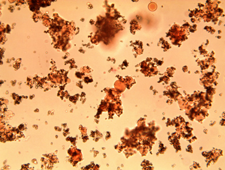
Figure 3. La3+ (150 µM)-induced fusion of erythrocytes of patients with diabetes. Incubation for 120 min at 37˚C × 100.
erythrocytes. Incubation of aggregates normal erythrocytes for 120 min at 37˚C does not cause cell fusion (association of contents cell) (Figure 1).
Figure 2 shows that La3+ induced extensive fusion of erythrocytes after 7 days of blood storage.