Synthesis, Characterization and Anticancer Activity Studies of New Schiff Base Pt (II) Complex ()
1. Introduction
Schiff bases play an important role in development of coordination chemistry. Many coordination compounds of biologically active ligands have synthesized and received much attention [1] [2] [3] .
Chelation causes drastic change in the biological properties or the ligands and also the metal moieties [4] . Azomethines exhibit a wide range of pharmacological activities like antimicrobial [5] , anti-parasitic [6] , anti-inflammatory [7] and anticancer [8] . Schiff base and its metal complexes possess several interesting biological activities such as anti-microbial and antitumor [9] - [15] .
In this work, Schiff base ligand was prepared and complexed with Potassium tetrarchloroplatinate (K2PtCl4). Characterization, electronic properties and anticancer effect are reported.
2. Experimental
2.1. Chemicals
The drug, chemicals and solvents used in this study were of analytical grade and used as obtained from Aldrich without further purification: Ibuprofen drug, hydrazine hydrate, Potassium tetrachloroplatinate (II) (K2PtCl4), Methanol (CH3OH), deionized water.
2.2. Instrumental
The melting points were measured on an electro thermal melting point apparatus and were not corrected. Fourier-transform infrared spectra were recorded using the KBr disc technique on a JASCO 410 FTIR spectrophotometer. Elemental (CHN) analysis was performed using an Exeter CE-440 elemental analyzer. UV-visible absorption spectra were measured in DMF (≈ 10−5 mole−1) using a Pye-Unicam 8800a uv-visible automatic scanning spectrophotometer.1 HNMR spectra of the ligand and its complex were recorded on a Varian Gemini-200 spectrometer (300 MHZ) using DMSO-d6 as solvent and TMS as internal reference. 13C NMR spectra of the ligand in DMSO was obtained using a Bruker 500 MHZ instrument using TMS as internal reference. Anticancer activity was evaluated at the Regional Center for Mycology and Biotechnology, Al-Azhar University, Cairo, Egypt.
2.3. Synthesis of Schiff Base
Synthesis of Schiff base L
The Schiff base ligand was prepared by condensation of mixture (0.001 mol) of 2-(4-isobutylphenyl)propanehydrazide and the 2,3,4-trihydroxybenzaldehyde in (30 ml) methanol. The mixture was refluxing for 24 h, The precipitated was filtered, crystallized by methanol, then dried in air for 24 h. Compound 2-(4-isobutylphenyl)propanehydrazide was prepared according the literature [16] .
Synthesis of Schiff base Platinum Complex (L-Pt)
The Schiff base Platinum complex (L-Pt) was prepared by mixing (0.001 mol) of K2PtCl4 was dissolved in 10 ml of methanol with some amount of deionized water and (0.001 mol) of Schiff base L in 20 ml of methanol. The mixture was refluxing for 24 h, The precipitated complex was filtered, washed twice with methanol, then dried in air for 24 h.
2.4. Cytotoxicity
Cell line Propagation
The cells were grown on RPMI-1640 medium supplemented with 10% inactivated fetal calf serum and 50 µg/ml gentamycin. The cells were maintained at 37˚C in a humidified atmosphere with 5% CO2 and were subculture two to three times a week.
Cytotoxicity evaluation using viability assay
For antitumor assays, the growth cell lines were suspended in medium at concentration 5 × 10⁴ cell/well in Corning® 96-well tissue culture plates, then incubated for twenty-four hour. The tested compounds were then additional into 96-well plates (3 replicates) to attain 12 concentrations for further compound. 6 vehicle controls with media or ½% dimethyl sulfoxide were run for each 96 well plate as a control. After incubating for twenty-four hour, the numbers of viable cells were determined by the MTT check. Briefly, the media was far from the 96 well plate and replaced with 100 µl of fresh culture RPMI 1640 medium without phenol red then ten µl of the twelve mM MTT, stock solution (five mg of MTT in one mL of PBS) to each well including the untreated controls. The 96 well plates were then incubated at 37˚C and five % CO2 for four hours. An 85 µl aliquot of the media was removed from the wells, and 50 µl of DMSO was added to each well and mixed thoroughly with the pipette and incubated at 37˚C for 10 min. Then, the optical density was measured at 590 nm with the microplate reader (SunRise, TECAN, Inc, USA)to see the quantity of viable cells and the percentage of viability was calculated as [(ODt/ODc)] × 100% where ODt is the mean optical density of wells treated with the tested sample and ODc is the mean optical density of untreated cells. The relation between living cells and drug concentration is plotted to get the survival curve of each tumor cell line after treatment with the required compound. The 50% inhibitory concentration (IC50), the concentration needed to cause toxic effects in 50% of intact cells, was calculable from graphic plots of the dose response curve for every concentration using Graph pad Prism software system (San Diego, CA. USA) [17] [18] .
3. Result and Discussion
3.1. Synthesis and Characterization
The Schiff base ligand was prepared by condensation of mixture (0.001 mol) of 2-(4-isobutylphenyl)propane hydrazide and the 2,3,4-trihydroxybenzaldehyde in (30 ml) methanol. The mixture was refluxing for 24 h, The precipitated was filtered, crystallized by methanol, then dried in air for 24 h. The Schiff base Platinum complex (L-Pt) was prepared by mixing (0.001 mol) of K2PtCl4 was dissolved in 10 ml of methanol with some amount of deionized water and (0.001 mol) of Schiff base L in 20 ml of methanol. The mixture was refluxing for 24 h, The precipitated complex was filtered, washed twice with methanol, then dried in air for 24 h. Table 1 summarized the physical properties (melting point, color, percentage yield, and elements analysis) of the ligand and its Pt complex. The proposed structure of Schiff base and L-Pt Complex were shown in Scheme 1.
![]()
Table 1. Physical properties of Schiff base and its complex.
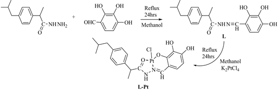
Scheme 1. The proposed structure of Schiff base L and its Pt complex L-Pt.
3.2. IR Spectra
The main IR Spectra of ligand and its complex are summarized in Table 2, The absence of the aldehydic carbonyl stretch in the ligand and the appearance of the characteristic azomethine υ C=N at 1633 cm−1 confirmed formation of the Schiff base. Comparison of infrared spectral data of Pt-complex and its corresponding ligand confirmed complexation as signification shifts in υ C=N was observed the strong band at 1633 cm−1 assigned to υ C=N in the free ligand shifted to higher wave number in Pt-complex at 1652 cm−1, indicating participation of azomethine nitrogen coordination. More ever, the single band at 3447 cm−1 due to the υ OH vibration was shifted upon complexation, which confirmed the involvement of the OH hydroxyl group in coordination to metal ions [19] [20] .
3.3. NMR Spectra
The main 1HNMR spectrum of the L, showed the azomethine proton at δ 8.6 ppm (s, 1H), while the multiples at δ (6.2 - 7.3) ppm (m, 6H) were ascribed to aromatic protons, the OH were observed at δ 8.8 ppm (s, 1H), 9.7 ppm (s, 1H) and 11.3 ppm (s, 1H).
For the 1HNMR spectrum of the Pt (II) complex an electron density shift was observed from ligand to metal signal of azomethine proton appeared at δ 7.5 ppm (s, 1H) as compared to 8.6 ppm in the ligand confirming coordination through the nitrogen of azomethine.
The 13C NMR spectrum of the Schiff base had characteristic signals at δ149 ppm of the azomethine carbon, C=N spectra were further characterized by absence of the aldehydic signal at δ 190 ppm from the corresponding aromatic aldehyde (Figure 1).
![]()
Table 2. Main IR absorption bands of Schiff base and its complex.
![]()
Figure 1. 1HNMR spectrum of the ligand L.
3.4. Electronic Spectra
Electronic spectra were recorded in DMSO. The UV-Vis peaks confined to π-π* and n-π* transitions of benzene and phenolic groups in the ligand. In the Schiff base, the band at 446 nm was attributed to the π-π *of the azomethine. Bands between 371 and 231 nm are associated with phenyl and amide π-π * transitions. In the spectra of the complexes, the π-π * of the azomethine shifted to a shorter wavelength observed at 434 nm, indicating that the hydrazone nitrogen was involved in coordination. The electronic spectra of the Pt (II) complexes had bands in the range of 381 - 227 nm shifted toward shorter and longer wavelength due to the n-π * and π-π * transitions of phenyl, lactam, and azomethine. In the spectra of the complex, the less intense and broad bands in the range of 580 - 660 and 757 nm resulted from the overlap of the energy π → π * transitions, mainly localized within the azomethine group, lactam and the LMCT transitions from the lone pairs of the phenolate oxygen and of the azomethane nitrogen donor to Pt (II) [21] [22] [23] .
![]()
Table 3. IC50 values (μg/ml) for Schiff base and Pt (II) complex tested against Helacells and PC3 cells.
3.5. Anticancer Activity
The results of in vitro anticancer activity of the tested the ligand and its Ptcomplex were evaluated for cytotoxicity against PC3 cells and Hela cells of humans in comparison with Cis palatine as a positive control. The L-Pt complex showed a higher cytotoxic activity than free ligand towards Hela cells and PC3 cells. Table 3 represents the cytotoxic activity of the tested compounds.
4. Conclusion
The present work describes the synthesis and in vitro anticancer activity of Schiff base and its complex Pt (II). The L-Pt complex showed a higher cytotoxic activity than Schiff base free ligand towards Hela cells and PC3 cells.