Immunohistochemical analysis of 8 biomarkers on tissue microarray (TMA) of 46 Moroccan invasive breast carcinoma ()
1. INTRODUCTION
Tissue Microarray (TMA) allows analysis of hundreds of specimens with one slide instead of incubating and analyzing samples one slide at a time. All the histochemical and molecular detection techniques that can be used with regular sections can also be used with tissue microarrays. Typical applications include immunohistochemical detection of protein expression of tumor specimens. It is also a tool in routine practice for quality control of immunohistochemistry techniques especially for hormone receptors and Her2/neu.
To introduce this technique in our institution, we chose to apply it to a set of Moroccan invasive breast carcinomas and study the immunohistochemical expression of 8 biomarkers of diagnostic, therapeutic and prognostic interest. Among the multiple markers reported in the literature, we chose to study the protein expression of oestrogen (ER) and progesterone (PR) hormone receptors, Her2/neu, 2 suppressor genes PTEN and p53, 2 proteins of the Wnt signalling pathway E-cadherin and beta-catenin and CK5/6.
2. MATERIAL AND METHODS
2.1. Specimen Selection
The paraffin blocs and hematoxylin-eosin stained slides of 46 cases of breast cancer were retrieved from the archives of the department of pathology of the Institute National d’Oncologie in Rabat, Morocco. Histologic types and Scarff and Bloom (SBR) grades after review were as shown in Table 1.
2.2. Tissue Microarrays (TMA) Construction
Areas of interest were selected on the slides and matched with the corresponding blocs Figure 1.
Three (3) cores, 1 mm in diameter each, were punched from each case and 2 receiver paraffin blocks were prepared with the Beecher manual arrayer (ATA-27; Beecher Instruments, Sun Prairie, WI). To orientate the microscopic analysis of the TMA slides the first spot was punched in a lung specimen and the last raw had less spots then the other raws.
The 2 TMA paraffin blocs were incubated for 30 mn at 37˚C and refrigerated during 30 mn.
Two hundred (200) five microns microtome sections were mounted on silanizated slides. A hematoxylin-eosin stain was performed every 40 sections to make sure that every case is at least represented by one spot. The slides are archived for potential new biomarkers analysis.

Table 1. Histologic types and grade.
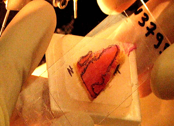
Figure 1. Normal and tumoral tissues delimited on the slide and the corresponding bloc with a permanent marker.
For our study, two slides were stained with the routine haematoxylin-eosin (HE) stain for morphologic analysis and 16 TMA slides were used for the immunohistochemical (IHC) analysis of the 8 biomarkers Figure 2.
2.3. Immunohistochemical Analysis
Immunohistochemical analysis was performed with the peroxydase-anti peroxydase technique and the 8 biomarkers studied were as shown in Table 2. The incubation time was 40 minutes for each biomarker.
2.4. Scoring of Biomarkers Staining Figure 3
Estrogen (ER) and Progesterone Receptors (PR): Nuclear immunostaining was scored negative if less than 10% tumour cells were immunostaining positive. Staining intensity was scored weak, moderate or strong.
Her2/neu: Membrane staining is scored 0: negative when no staining is observed or membrane staining is less than 10% of tumour cells, 1+: negative when a faint/barely perceptible membrane staining is detected in more than 10% of the tumour cells and the cells are only stained in part of the membrane, 2+: positive when a weak to moderate complete membrane staining is ob-

Table 2. Characteristics of the antibodies used in this study.
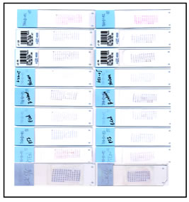

Figure 2. TMAs of the 46 cases of invasive breast carcinoma.

Figure 3. Hematoxyline-eosin stain [1] Grade 3 [2] Grade 2, Hormone Receptors [3], HER2/neu [4], Betacatenin [5], E-cadherin [6], p53 [7] and PTEN [8].
served in more than 10% of the tumour cells and 3+: positive when a strong complete staining is observed in more than 30% of the tumour cells.
E-cadherin and beta-catenin: A histochemical score (H-score) was used and included an assessment of both the intensity of staining and the percentage of stained cells. For the intensity, a score index of 0, 1, 2 and 3 corresponding to negative, weak, moderate and strong staining intensity was used and the percentage of positive cells at each intensity was estimated subjectively. A final score of 0 - 300 is the product of both the intensity and the percentage.
Reduced expression was considered when H-score was equal or less than 100. We took into account all the structures stained (C: cytoplasm, C/M: cytoplasm and membrane, C/M/N: cytoplasm and /or membrane and nucleus)
p53: Nuclear immunostaining for p53 was scored negative if less than 10% tumour cells were immunostaining positive, + for 30% - 60%, ++ for 60% - 90% and +++ if more than 90%. Staining intensity was scored weak, moderate or strong.
PTEN: The immunohistochemical expression of breast cancer cells was judged to be either normal, reduced or lost compared with the PTEN protein expression of the normal mammary gland. Staining intensity is assigned as 0, negative; 1, weak; 2, moderate; 3, strong. Percentage of positive cells is defined as 0: less than 1%; 1: 1% - 10%; 2: 11% - 50%; 3: 51% - 80%; and 4 more than 80% positive cells.
Ck5/6: any cytoplasmic expression in definite neoplastic cells was considered to be positive.
3. RESULTS
Only 18 slides had to be analyzed instead of 370 slides if each case had to be represented on a separate slide.
The results of biomarker expression according to histologic grade and phenotype are summarized in Tables 3 and 4.
3.1. Hormones Receptors (HR), Her2/neu and CK 5/6 Results In our sudy, 58.69% of the cases (27/46; all 4 ILC were positives) were ER positive of which 62.96% (17/27) had

Table 3. Expression of the 8 biomarkers correlated to tumour grade of IDC and ILC.

Table 4. Biomarker expression of the 19 triple negative (HR and Her2/neu negative) IDC.
PR concomitant expression. ER positive cases were IDC grade 2 in 82.35% of the cases (14/17) and grade 3 in only 27.77% (5/18) of the cases. ER negative cases were IDC grade 3 in 72.22 % (13/18).
We could not look for gene amplification by FISH or CISH for the two Her2/neu 2+ cases; all the remaining 44 cases were HER2/neu negative. 43.18% (19/44) were triple negative breast cancers (TNBC) defined as HR and Her2/neu negative of which 15.78% (3/19) were of the basal phenotype expressing CK5/6. On the other hand, 72.22% (13/18) of the TNBC cases were IDC grade 3. Of the 18 IDC grade 3, 22.22% (4/18) were CK5/6 positive.
3.2. E-Cadherin and Beta-Catenin
Reduced membranous expression was observed in 41.30% (19/46) of the cases for E-cadherin and only 10.86% (5/46) of the cases for beta-catenin with concomitant reduction in 3 cases.
6.52% (3/46) of the cases showed loss of E-cadherin expression with concomitant loss of beta-catenin expression in one case.
Among IDCs, 37.5% (15/40) had reduced expression of E-cadherin.
Cytoplasmic staining for beta-catenin was observed in 97.82% (45/46, 1 colloid carcinoma was negative) with concomitant beta-catenin nuclear staining in 20% (9/45) of the cases (1 ILC grade 2, 1 invasive colloid carcinoma and 7 IDC grade 2).
3.3. P53 and PTEN
PTEN expression was reduced in 76.08% (35/46) and lost in 10.86% (5/46) among which 3 were IDC/TNBC.
p53 was over-expressed in 10.86% (5/46) of the cases (5/46; 2 IDC grade 2, 2 IDC grade 3 triple negative and 1 ILC grade 2).
4. DISCUSSION
Breast cancer is the leading cancer in women worldwide. In Morocco, the incidence was 39, 9 new cases /100 000 women in 2007 and represents 1/3 of all cancers in women [1].
Diagnostic, prognostic and predictive factors rely on morphological parameters but also on molecular and genetic biomarkers with the aim of a personalized treatment according to the patient’s tumor profile.
Therefore, TMA is the best choice to test new biomarkers on big sets of tumors with cost and time savings.
In our study, we only had to analyze 2 HE and 16 IHC slides instead of 370 slides which illustrates the economic advantages of this technique but also the comfort and the facility for the pathologist to do comparative analysis on the same slide.
4.1. Hormone Receptors
Estrogen Receptor (ER) status is considered a predictive factor for breast cancer response to hormonal treatment, ER+ tumors being more responsive than ER− tumors. Moreover, ER+ tumors have better survival than ERtumors and ER seems to define sub-groups with different tumor characteristics which might be modified by PR status.
Several studies report that 50% to 85% of breast carcinomas are ER+ and PR being less frequently positive when ER is negative (5% of the cases) [2]. None of our cases was PR positive and ER negative.
13/19 ER negative cases were grade 3 IDC. This is in accordance with the data in the literature and doesn’t seem to vary by racial/ethnic group [3].
On the other hand, it was reported that ER might suppress p53 function by binding directly to p53 and that ionizing radiation interrupt this interaction. It is therefore important to be aware of the tumor ER and p53 status when combining anti-ER and radiation therapy. In our study, 3 cases only were p53 + and ER+ [4].
Colloide carcinomas are good prognostic tumors often ER positive [2]. Our case was both ER and PR positive.
4.2. HER2/neu
HER2/neu amplification determined by FISH (Fluorescent in situ Hybridization) or CISH (Chromogenic in situ hybridization) or Her2/neu overexpression determined by immunohistochemistry seems to occur in 10% - 30% of breast cancer and is considered a marker of poor prognosis as well as an indicator of response to Trastuzumab, an anti-Her2 antibody, in metastatic breast cancer [2,5-9].
On the other hand, HER2/neu positive patients develop chemoresistance when given adjuvant chemotherapy (cyclophosphamide, methotrexate, and fluorouracil) [8,9] whereas in a neoadjuvant situation some authors report a 50% decrease in relapse risk.
Response to hormone therapy is controversial with a bad response of HER2/neu positive tumors to Tamoxifene [6].
Except for the 2 cases which we could not analyze by FISH or CISH to look for the presence or absence of amplification, all the 44 cases were negative.
41.30% (19/46) of our cases were HR and HER2/neu negatives a so called triple-negative (TNBC) group of high risk breast cancer which cannot benefit from specific therapies, such as (Trastuzumab and Tamoxifene) targeting these proteins. 13/19 of these cases were grade 3 IDC and this is in accordance with the data in the literature [10]. In this study, there was also a positive expression of basal cytokeratins and it was suggested to use the basal phenotype together with androgen receptor status and other pathologic variables (lymph node status and tumor size) to select high-risk and low-risk patients with the possibility of treatment options in this high risk group.
Our results with CK5/6 are similar to those reported in the literature, 7.7% to 38.5% of breast carcinomas being reported with a basal phenotype [11]. Other authors consider [5-13], grade 3 IDC as a heterogenous group of tumors and those with a basal phenotype having a shorter disease-free and overall survival and less response to standard adjuvant chemotherapy [14]with indication of new therapeutic approaches.
Nevertheless, this sub-group could benefit from antiEGFR targeted therapy, taxanes and anti-PARP (poly (ADP-ribose) polymerase) molecules [15].
For Piekarski et al. the basal phenotype has a propensity for distant metastasis and is characterized by HR negativity, p53 positivity and reduced expression of PTEN [11].
4.3. E-Cadherin
E-cadherin is a cell adhesion molecule considered as a suppressor gene expressed in normal breast tissue but is often irreversibly absent in lobular carcinoma thus its use as a phenotypic marker in breast cancer diagnosis.
In the more common ductal carcinoma, variable Ecadherin expression is reported in 50% of the cases [16].
The data in the literature associate reduced E-cadherin expression with an increased risk of the development of distant metastasis and local and regional recurrence and its assessment by immunohistochemistry is recommended to help in the identification of patients with poor outcomes and as an indicator of disease-free interval [17].
4.4. BETA-Catenin
Beta-catenin is reported to exist at the cell membrane as part of the adherens complex with E-cadherin. Its expression in the cell cytoplasm and nucleus has a different prognostic value according to its localization [18].
Cytoplasmic expression of beta-catenin seems to correlate with a favourable tumor phenotype with prolonged disease-free and overall survival whereas nuclear expression of beta-catenin seems to indicate and aggressive tumor phenotype negatively affecting overall survival [18].
Several mechanisms may induce beta-catenin accumulation but it was found that beta-catenin mutations are rare in breast cancer and that other proteins involved in the Wnt signalling pathway or independent of the Wnt pathway may lead to beta-catenin accumulation as it is the case when PTEN and p53 suppressive functions are lost [19,20].
Our results are much higher than the 60% reported in the literature [20].
4.5. PTEN
PTEN is a tumor-suppressor gene and its inactivation, by loss of heterozygosity or mutation, with decreased PTEN expression is correlated with breast cancer invasiveness, lymph node metastasis and a worse prognosis [21]. In our study, PTEN reduced expression was observed in 76.08% (35/46) of our cases which is much higher than the 38% reported in Bose’s study and the 33% reported by Perren [22]. This variability might also be the consequence of different antibodies used in IHC analysis.
In the same studies, PTEN loss was reported in association with negative estrogen and progesterone receptor and in our study PTEN loss was found in 10.86% (5/46) of our cases of which 3 cases were both ER and PR negatives [22]. Bose also reported PTEN loss in 11% of in situ breast carcinoma suggesting that PTEN loss might be an early step in breast carcinogenesis.
On the other hand, loss of PTEN was reported to predict Trastuzumab resistance in vitro, in vivo and in the clinical setting [23]. This resistance is reversible with the adjunction of P13K inhibitors.
In a multivariate analysis, Lee et al. [24] attested the prognostic value of PTEN reduced expression with the proposal of introducing PTEN evaluation in routine practice.
4.6. p53
p53 mutation is reported to be one of the most common mutations in human cancer the frequency of PTEN mutation approaching that of p53 [23]. In our study, p53 was overexpressed in 10.86%. We did not carry genetic analysis on our cases, but our results might be explained by false-negative results since it has been reported that tumors containing homozygous deletions or non-sense and truncating mutations might not lead to p53 protein accumulation detectable by immunohistochemistry [24].
Although alterations in p53 function has been reported to correlate with response to chemotherapeutic agents [13-25] data on its usefulness as a clinical prognostic factor in breast cancer are controversial some authors reporting p53 as an indicator of poor prognosis [15] while others find no correlation with survival [26].
p53 statut has less prognostic significance compared with the usual prognostic factors [26,27].
5. CONCLUSIONS
The aim of our study was to introduce TMA technique in our hospital which is considering a reference institution for cancer in morocco. We chose to apply the technique to 46 cases of invasive breast carcinomas. Although no statistical study was performed to look for any significance of the results obtained, we found good correlation with the data in the literature for ER positivity and tumor grade, ER negative tumors been mostly of grade 3. For the triple-negative tumors (HR and Her2/neu negative) we also found the correlation reported in the literature with grade 3 tumors, basal phenotype and loss of PTEN.
E-cadherin was mostly reduced in ductal carcinoma with loss in only one case.
All tumors but one expressed cytoplasmic beta-catenin (with concomitant nuclear expression in some cases) which is higher than the data in the literature and seems to correlate with poor prognosis.
PTEN expression was reduced or lost in a much higher proportion than p53.
Several mechanisms are reported to interact to produce tumor development and progression, but in our study beta-catenin cytoplasmic expression seems to correlate most with the data in the literature stating that beta-catenin can be involved in breast cancer formation and/or progression and may serve as a target for breast cancer therapy and Wnt pathway thus offering targeting points for new cancer drug approaches.
TMA is a feasible tool to study a large number of cases allowing comparative analysis of the expression of different biomarkers. To our knowledge, this is the first study of 8 biomarkers to be done on TMAs from 46 moroccan invasive breast carcinomas. This would allow for larger studies with the aim of analysing the significance of these biological markers and their correlation with clinicopathologic variables and their impact in personalized therapeutic decisions.
6. ACKNOWLEDGMENT
The authors would like to warmly thank INO staff, particularly Boudaz Khalid and Bathami El Khayati for samples collection, the Department of Pathology Core Facility staff at Columbia University Medical Center (New York, USA), particularly Lyn Yang and Rina Wu for their excellent technical work and the International Agency of Atomic Energy, (Wien, Austria) for financing the study.
NOTES