Management of Epidermoid Carcinoma of the Conjunctiva in a Melanoderma Subject in a Secondary Ophtalmology Centre: About a Case ()
1. Introduction
Conjunctival squamous cell carcinoma (CSC) is a malignant tumour of limbal stem cells. It is the invasive stage of squamous neoplasia of the ocular surface with the invasion of the epithelial basement membrane and the lamina propria. Its clinical distinction from other amelanotic tumours is based on certain clinical and especially histological features [1]. Typically, the lesions present as sessile, white or pink “cauliflower” tumours, slightly elevated, or budding, most often with ulcerations, and sometimes ruptured on the surface. Although they have a limited relationship with the surrounding healthy tissue, they are accompanied by typical vascularity of multiple capillaries that form the central vascular axis of the lobules and terminate in the glomerular coves, giving them a raspberry appearance [1]. It usually starts from the conjunctiva between the eyelids and then grows and overlaps with the conjunctiva of the eyelid margins. It may or may not invade the cornea [1]. Squamous cell carcinoma is larger and thicker than carcinoma in situ and is more or less covered with keratin. The non-keratinised type must be differentiated clinically from benign papilloma or carcinoma in situ. The bulbar conjunctiva is the most common location, but lesions can form in the fornix of the palpebral conjunctiva and anterior conjunctiva, and in the caruncles. CSC can exist in different clinical forms [1]: the gelatinous form is the most common, and the nodular type develops rapidly, which is associated with a high risk of lymph node metastasis. The diffuse type is usually difficult to diagnose and is similar to chronic conjunctivitis [1]. The leukoplakic type, which raises the question of differential diagnosis with non-invasive OSNN (Ocular Surface Squamous Neoplasia). Mainly the extension of CSC is local, more superficial than deep. It is referred to as horizontal and vertical progression, which can go from the “tree root” to the sclera, explaining the difficulty of complete deep resection and the high frequency of tumour recurrence.
It represents 5% of all ocular malignancies [2]. It has an incidence of 8.4 per million population [3]. It is a tumour that develops in the elderly population, but younger cases can occur in subjects with Xeroderma pigmentosum [4]. The male sex is most often affected [5] and frequently seen in fair-skinned subjects than in pigmented subjects [6].
We report the case of a 44-year-old female patient with unilateral budding squamous cell carcinoma.
2. Patient and Observations
The patient was 44 years old and consulted for the first time for a swelling that had been evolving for 3 months associated with redness, ocular irritation and foreign body sensations.
The ophthalmological examination revealed a visual acuity of 10/10 in both eyes and revealed a nodular, budding, pigmented, vascularised temporal tumour mass in the right eye, measuring approximately 6 mm in diameter, mobile, no adherent to the deep plane and purely conjunctival (Picture 1: aspect of the tumour from the front and the side). The rest of the anterior segment and the fundus were normal.
All lymph nodes were free, with no pre-tragic or sub-maxillary adenopathy.
The extension work-up, human immunodeficiency virus (HIV) serology and human papilloma virus (HPV) were negative. Magnetic resonance imaging (MRI) and computed tomography (CT) were not available.
Due to the size and appearance of the tumour, a nodular pigmented squamous
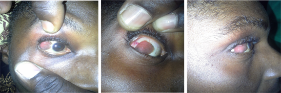
Picture 1. Appearance of the tumor from the front and profile.
cell carcinoma of the conjunctiva was suspected.
Simple excision of the tumour was performed with a healthy margin of about 3 to 4 mm without adjuvant treatment not available in our secondary ophthalmology centre.
The choice of treatment was motivated by the absence of adjuvant therapies (cryotherapy, radiotherapy, chemotherapy and topical/general immunotherapy), the predictive factors of recurrence which are: advanced age, histological type, positive excision margins, tumour location and lesion size (greater than 10 mm). Adjuvant therapy is intended to enhance the success of surgery.
Treatment with local corticosteroids and eyewash was initiated postoperatively.
An anatomopathological examination was requested and the histopathological report concluded an invasive squamous cell carcinoma.
The postoperative follow-up at 6 and 18 months was favourable with a normal appearance of the conjunctiva and transparency of the cornea (Picture 2).
This proves that in the absence of adjuvant treatments such as chemotherapy and others, factors predictive of recurrence and at an early stage that it is possible to perform a simple excision of conjunctival squamous cell carcinoma. However, given the malignant nature of the tumour, surveillance is always necessary.
3. Discussion
Classically, carcinomatous lesions present as a sessile, slightly elevated, vegetative or budding white or pinkish “cauliflower” tumour, often ulcerated and sometimes with a fissured surface. Well-limited in relation to the surrounding healthy tissue, they are accompanied by a typical vascularisation consisting of several capillaries that form the central vascular axis of the lobules and terminate in glomerular loops, giving them a raspberry-like appearance [6] [7]. CSC can present in different clinical forms [8]: the gelatinous form is the most common. The nodular form has a rapid course with a high risk of lymph node metastasis. The diffuse form is often difficult to diagnose [9]. The leukoplakic form poses the problem of differential diagnosis with non-invasive OSNN (Ocular Surface Squamous Neoplasia) [1].
Our case was a pigmented nodular form with a purely unilateral conjunctival

Picture 2. Appearance of the area of excision at 18 months.
location without extension.
Data from the African literature show that CSC is a common malignancy in the tropics. It represents 33.5% of all malignant tumours of the eye and adnexa [10]. In Ivory Cost, Bereté and al, reported 26 cases in 4 years [11]; in Martinique, 4 cases were reported in 5 years in a mixed population (3 melanoderma and 1 Caucasian) [12]. In Senegal Secka S. M. et al., found that it was the second most common ocular cancer with 15.56% after retinoblastoma and the first conjunctival tumour with 65.56% [13].
It is a tumour that develops in elderly subjects around the sixth decade, the youngest cases would have occurred in patients with xeroderma pigmentosum [4] [6], whose incidence is 75%.
The various risk factors listed are mainly ultraviolet exposure and immunosuppression [12].
The case we report was 44 years old, melanodermal, female, and could be explained by the combined action of exposure to solar radiation and dust.
Despite the young age of our patient, she was not found to be infected with HIV or HPV and had not been treated with immunosuppressants. Some authors believe that HIV and HPV infections play a role in the genesis of squamous cell carcinoma [14] [15]. Several cases of CSC have been described in HIV positive subjects. BERETE found that 76.92% of CSC cases were HIV positive with a statistically significant association [11]. In Kenya, the prevalence of CEC was 7.8% among HIV positive patients [16] and the average age of patients was 38.3 years. In Tanzania, in areas of the AIDS epidemic, an increase in the incidence of CSC was reported, with 10 cases in 1994 and 40 cases in 1997 [17].
When CSC occurs at early ages HIV infection should be investigated [18].
Cases have been reported in patients undergoing long-term immunosuppressive therapy for transplantation [19].
The treatment of CSC is mainly based on surgery, with or without other adjuvant therapies such as cryotherapy, radiotherapy, chemotherapy and topical and systemic immunotherapy. The aim of surgery is to remove the entire tumour with a safety margin of healthy tissue to avoid recurrence [1]. Adjuvant therapy is intended to enhance the success of surgery.
For invasive squamous cell carcinomas, excisional biopsy combined with cryotherapy is performed. If the excisional margins are healthy, simple surveillance is performed. If the margins are affected, topical chemotherapy with 0.04% mitomycin C and possibly radiotherapy are performed [1].
In our case, we performed a simple excision with a healthy margin of 3 to 4 mm without adjuvant treatment.
The postoperative follow-up was favourable at 6 and 18 months after tumour removal (Picture 2).
The choice of this technique was motivated by the absence of predictive factors for recurrence, which are: advanced age, histological type, positive margins of excision, location of the tumour and size of the lesion (greater than 10 mm).
4. Conclusions
Conjunctival squamous cell carcinoma is a common malignancy in tropical environments. Prolonged sun exposure is unanimously recognised as a predisposing factor. It most often affects elderly people, but can be seen in young people. This case demonstrates the predisposition of young subjects with pigmented skin but above all reinforces the hypothesis of exposure to sunlight as a favouring factor.
The anatomopathological study of the specimen is the only way to guarantee an early diagnosis of certainty. Early management would reduce recurrence and the need for adjuvant treatment such as cryotherapy, radiotherapy, chemotherapy and topical and general immunotherapy. These adjuvant treatments have a cost and are not always available in our countries.