A Rare Cause of Gastrointestinal Bleeding: Cholecystoenteric Fistula ()
1. Introduction
Acute upper gastrointestinal bleeding is a common medical emergency that could involve the vital prognosis. The etiologies are many: some are common, others are rare. Among the very rare etiologies, the fistulas whose diagnosis can be done by endoscopy and imaging and treatment is usually surgical.
2. Case Report
A 60-year-old woman presented to the emergency department of our university hospital for several episodes of upper gastro-intestinal bleeding (hematemesis and melena), the first of which dated to 3 days. On examination, hemodynamic parameters were correct. At admission, her haemoglobin was 6.9 g/dl, platelet count 213.000/mcl, prothrombin time 87% and liver test didn’t show any abnormality. The endoscopic examination (Figure 1) showed a large ulcer at the front of the bulb, with a calculation within it and a visible vessel on the banks of the ulcer treated by putting two clips to prevent bleeding reccurence.
Computer tomography (CT), demonstrated a sclerotic, atrophic and multi gallstone gallbladder, with a calculation protruding in the duodenum (Figure 2). Several mechanisms have been reported in the literature explaining
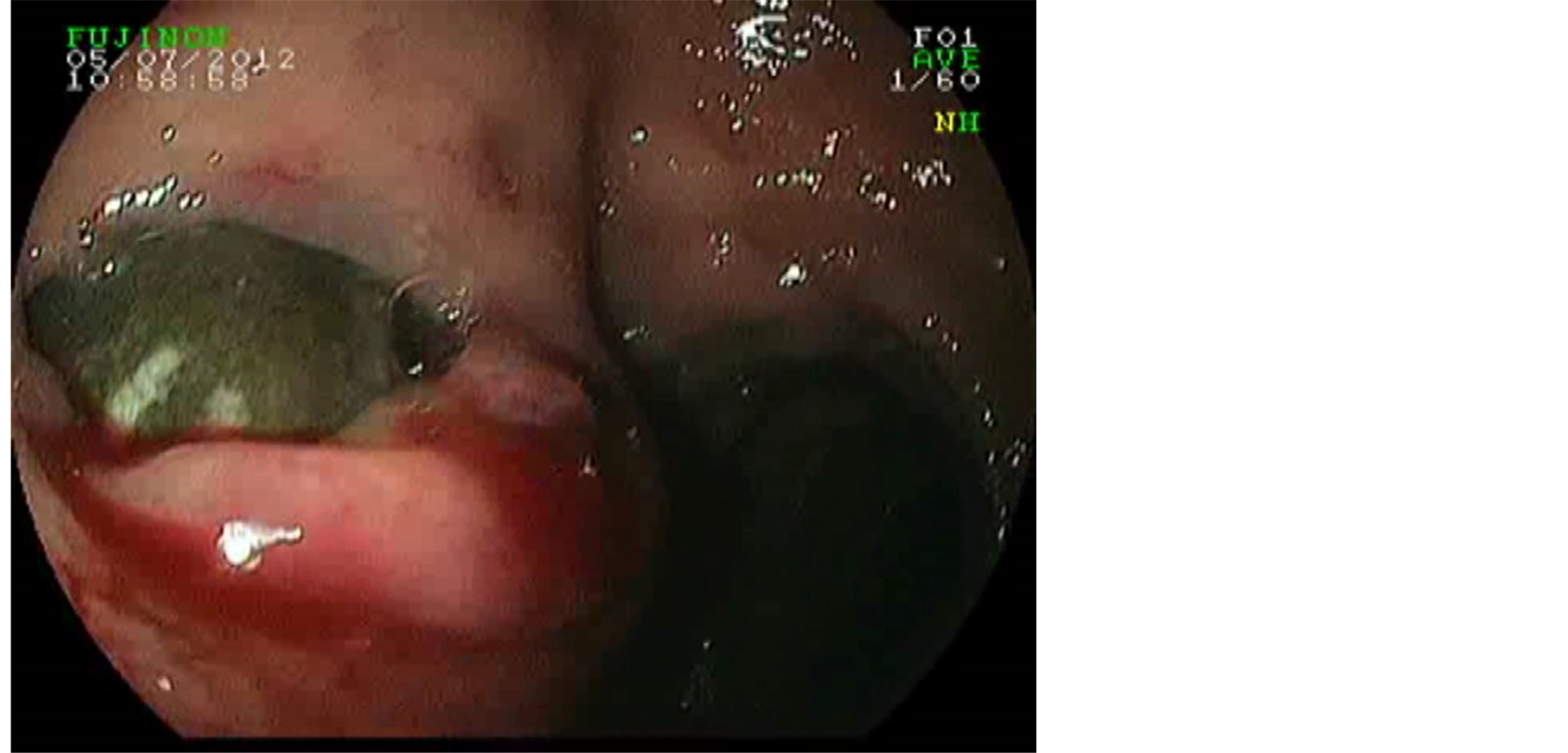
Figure 1. Large ulcer at the front of the bulb with a calculation within it.
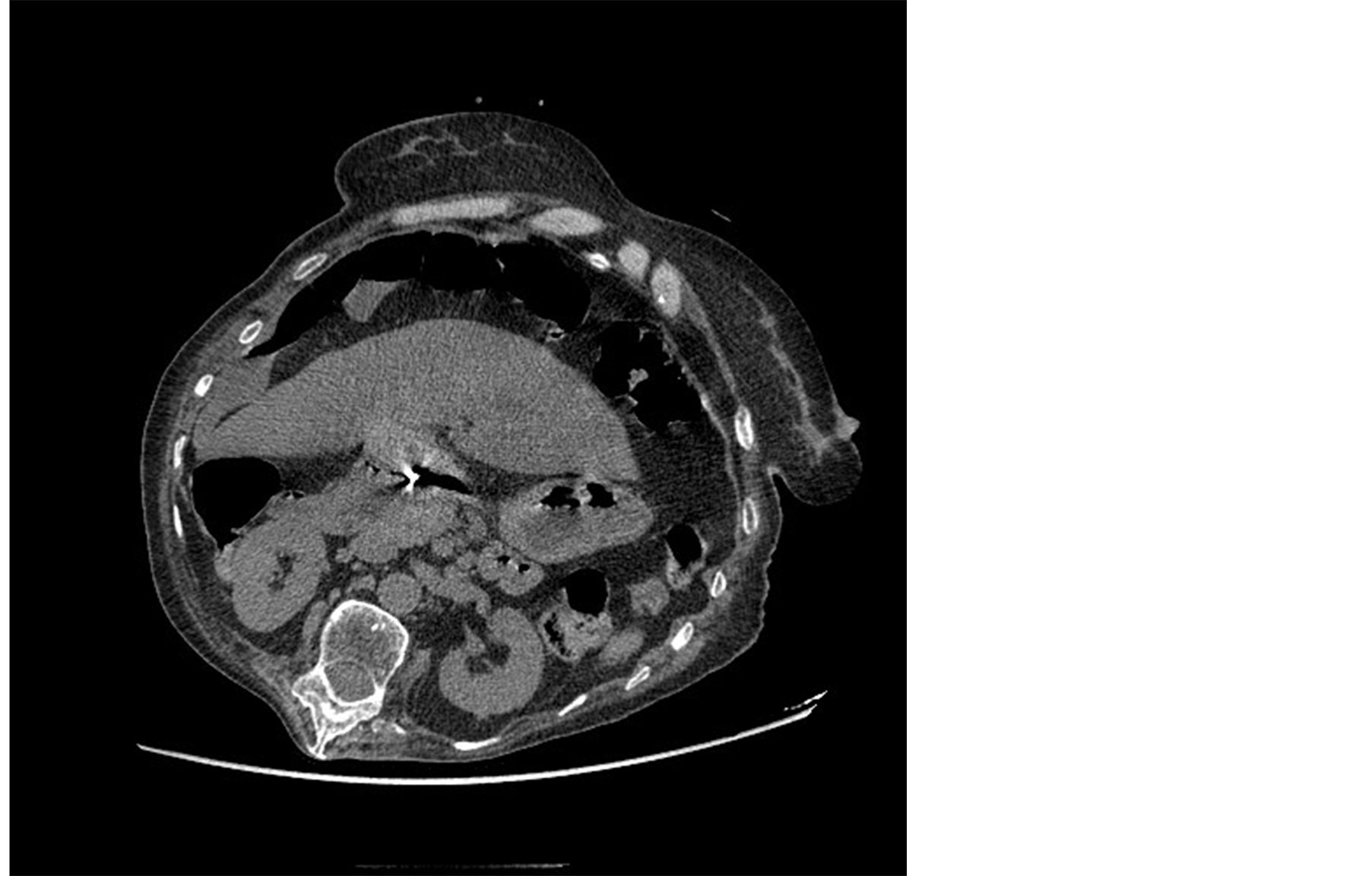
Figure 2. Sclerotic, atrophic gallbladder, with a calculation protruding in the duodenum.
the appearance of this kind of fistula, including pseudoaneurysm and haemobilia [1] -[3] . The patient underwent surgery which consisted on partial cholecystectomy with duodenal suture and closing of cystic duct.
3. Conclusion
Via this case, we want to attract attention to this rare cause of gastrointestinal bleeding, and we emphasize the importance of careful endoscopic examination in upper gastrointestinal bleeding to be able to reveal this kind of causes. CT performed by an expert can help in diagnosis. Surgery is the standard treatment for this cause of upper gastrointestinal bleeding.
4. Study Highlights
Bilio-digestive fistula as rare disease.
Bilio-digestive fistula as a rare cause of gastrointestinal bleeding.
The importance of careful endoscopy.
The importance of imaging (CT).
Surgery as the standard treatment.
NOTES
*Corresponding author.